Chlorine in PDB 1utr: Uteroglobin-Pcb Complex (Reduced Form)
Chlorine Binding Sites:
The binding sites of Chlorine atom in the Uteroglobin-Pcb Complex (Reduced Form)
(pdb code 1utr). This binding sites where shown within
5.0 Angstroms radius around Chlorine atom.
In total 4 binding sites of Chlorine where determined in the Uteroglobin-Pcb Complex (Reduced Form), PDB code: 1utr:
Jump to Chlorine binding site number: 1; 2; 3; 4;
In total 4 binding sites of Chlorine where determined in the Uteroglobin-Pcb Complex (Reduced Form), PDB code: 1utr:
Jump to Chlorine binding site number: 1; 2; 3; 4;
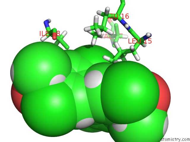
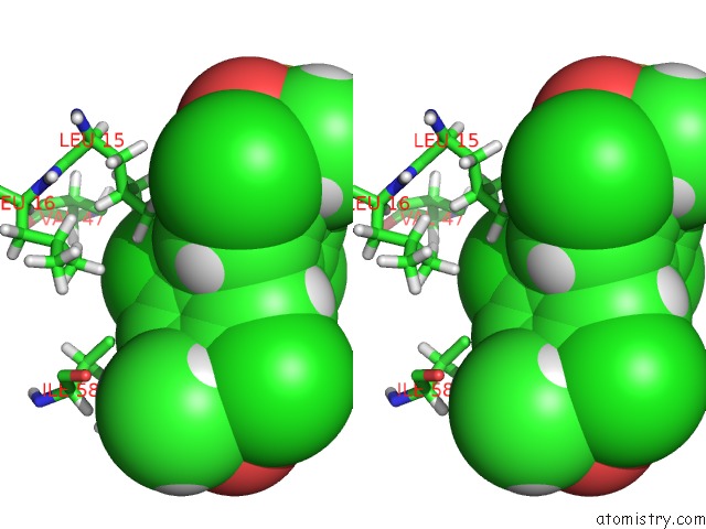
Chlorine binding site 1 out of 4 in 1utr
Go back to
Chlorine binding site 1 out
of 4 in the Uteroglobin-Pcb Complex (Reduced Form)
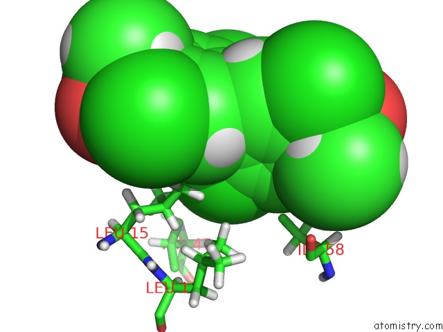
Mono view
Stereo pair view
Mono view
Stereo pair view
A full contact list of Chlorine with other atoms in the Cl binding
site number 1 of Uteroglobin-Pcb Complex (Reduced Form) within 5.0Å range:
|
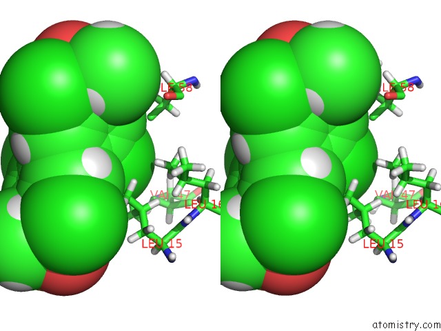
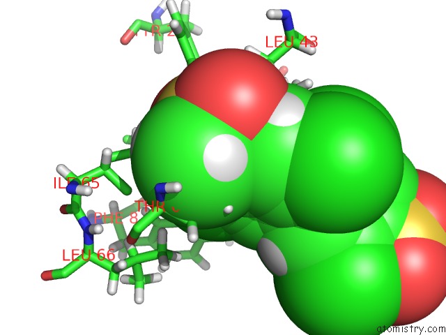
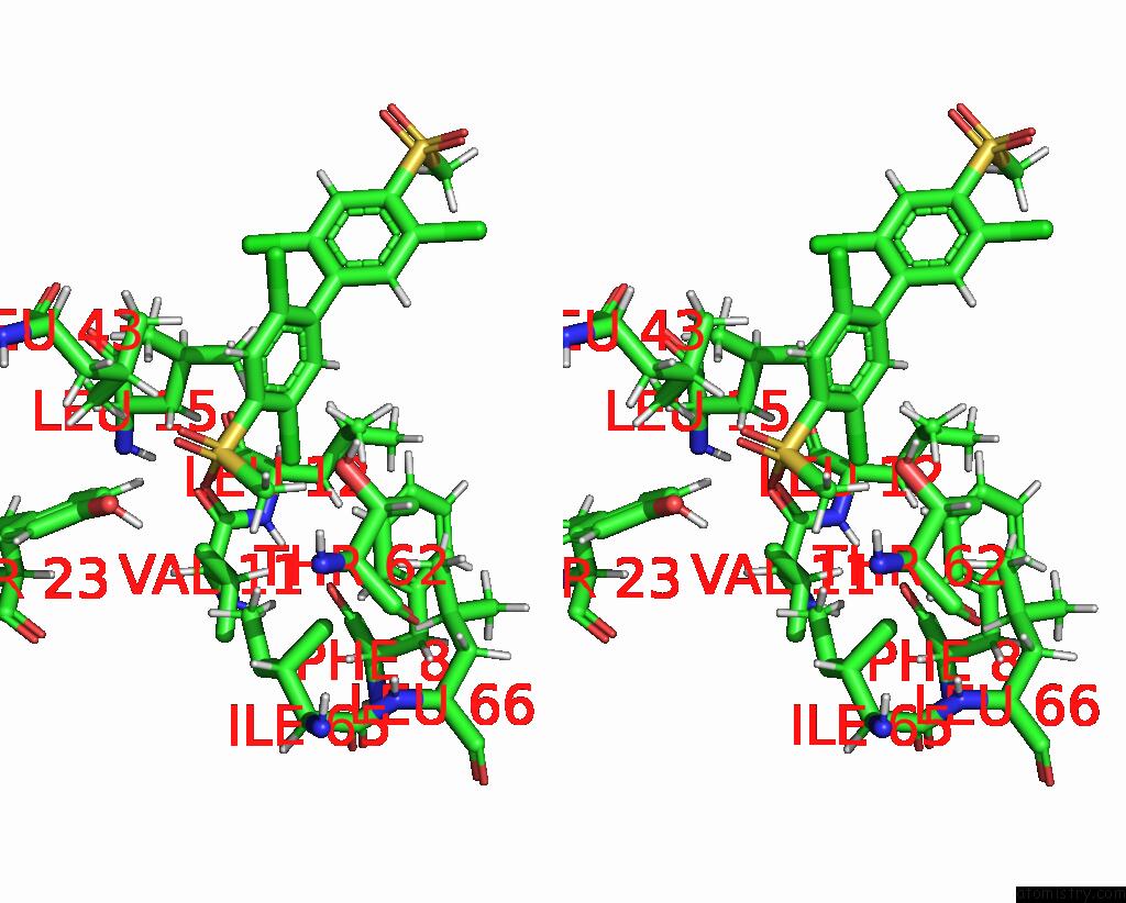
Chlorine binding site 2 out of 4 in 1utr
Go back to
Chlorine binding site 2 out
of 4 in the Uteroglobin-Pcb Complex (Reduced Form)
Mono view
Stereo pair view
Mono view
Stereo pair view
A full contact list of Chlorine with other atoms in the Cl binding
site number 2 of Uteroglobin-Pcb Complex (Reduced Form) within 5.0Å range:
|
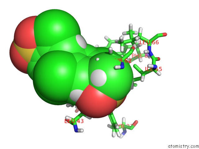
Chlorine binding site 3 out of 4 in 1utr
Go back to
Chlorine binding site 3 out
of 4 in the Uteroglobin-Pcb Complex (Reduced Form)
Mono view
Stereo pair view
Mono view
Stereo pair view
A full contact list of Chlorine with other atoms in the Cl binding
site number 3 of Uteroglobin-Pcb Complex (Reduced Form) within 5.0Å range:
|
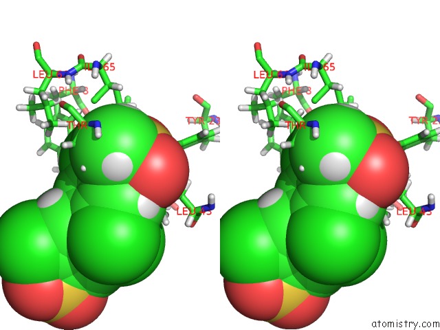
Chlorine binding site 4 out of 4 in 1utr
Go back to
Chlorine binding site 4 out
of 4 in the Uteroglobin-Pcb Complex (Reduced Form)
Mono view
Stereo pair view
Mono view
Stereo pair view
A full contact list of Chlorine with other atoms in the Cl binding
site number 4 of Uteroglobin-Pcb Complex (Reduced Form) within 5.0Å range:
|
Reference:
T.Hard,
H.J.Barnes,
C.Larsson,
J.A.Gustafsson,
J.Lund.
Solution Structure of A Mammalian Pcb-Binding Protein in Complex with A Pcb. Nat.Struct.Biol. V. 2 983 1995.
ISSN: ISSN 1072-8368
PubMed: 7583672
DOI: 10.1038/NSB1195-983
Page generated: Sat Jul 20 02:50:04 2024
ISSN: ISSN 1072-8368
PubMed: 7583672
DOI: 10.1038/NSB1195-983
Last articles
Zn in 9J0NZn in 9J0O
Zn in 9J0P
Zn in 9FJX
Zn in 9EKB
Zn in 9C0F
Zn in 9CAH
Zn in 9CH0
Zn in 9CH3
Zn in 9CH1