Chlorine »
PDB 4da0-4dhm »
4dab »
Chlorine in PDB 4dab: Crystal Structure of the Hexameric Purine Nucleoside Phosphorylase From Bacillus Subtilis in Complex with Hypoxanthine
Enzymatic activity of Crystal Structure of the Hexameric Purine Nucleoside Phosphorylase From Bacillus Subtilis in Complex with Hypoxanthine
All present enzymatic activity of Crystal Structure of the Hexameric Purine Nucleoside Phosphorylase From Bacillus Subtilis in Complex with Hypoxanthine:
2.4.2.1;
2.4.2.1;
Protein crystallography data
The structure of Crystal Structure of the Hexameric Purine Nucleoside Phosphorylase From Bacillus Subtilis in Complex with Hypoxanthine, PDB code: 4dab
was solved by
N.H.Martins,
P.O.Giuseppe,
A.N.Meza,
M.T.Murakami,
with X-Ray Crystallography technique. A brief refinement statistics is given in the table below:
| Resolution Low / High (Å) | 41.23 / 1.85 |
| Space group | P 63 2 2 |
| Cell size a, b, c (Å), α, β, γ (°) | 135.394, 135.394, 58.006, 90.00, 90.00, 120.00 |
| R / Rfree (%) | 20.1 / 24.1 |
Chlorine Binding Sites:
The binding sites of Chlorine atom in the Crystal Structure of the Hexameric Purine Nucleoside Phosphorylase From Bacillus Subtilis in Complex with Hypoxanthine
(pdb code 4dab). This binding sites where shown within
5.0 Angstroms radius around Chlorine atom.
In total 3 binding sites of Chlorine where determined in the Crystal Structure of the Hexameric Purine Nucleoside Phosphorylase From Bacillus Subtilis in Complex with Hypoxanthine, PDB code: 4dab:
Jump to Chlorine binding site number: 1; 2; 3;
In total 3 binding sites of Chlorine where determined in the Crystal Structure of the Hexameric Purine Nucleoside Phosphorylase From Bacillus Subtilis in Complex with Hypoxanthine, PDB code: 4dab:
Jump to Chlorine binding site number: 1; 2; 3;
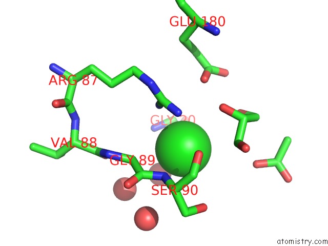
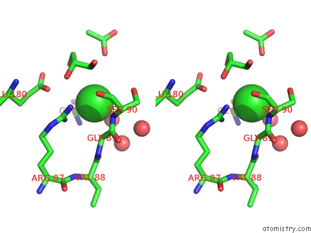
Chlorine binding site 1 out of 3 in 4dab
Go back to
Chlorine binding site 1 out
of 3 in the Crystal Structure of the Hexameric Purine Nucleoside Phosphorylase From Bacillus Subtilis in Complex with Hypoxanthine
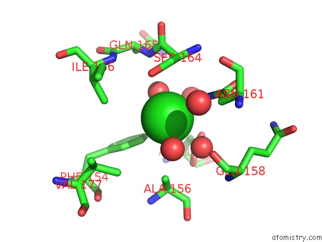
Mono view
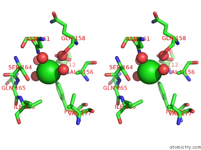
Stereo pair view
Mono view
Stereo pair view
A full contact list of Chlorine with other atoms in the Cl binding
site number 1 of Crystal Structure of the Hexameric Purine Nucleoside Phosphorylase From Bacillus Subtilis in Complex with Hypoxanthine within 5.0Å range:
|
Chlorine binding site 2 out of 3 in 4dab
Go back to
Chlorine binding site 2 out
of 3 in the Crystal Structure of the Hexameric Purine Nucleoside Phosphorylase From Bacillus Subtilis in Complex with Hypoxanthine
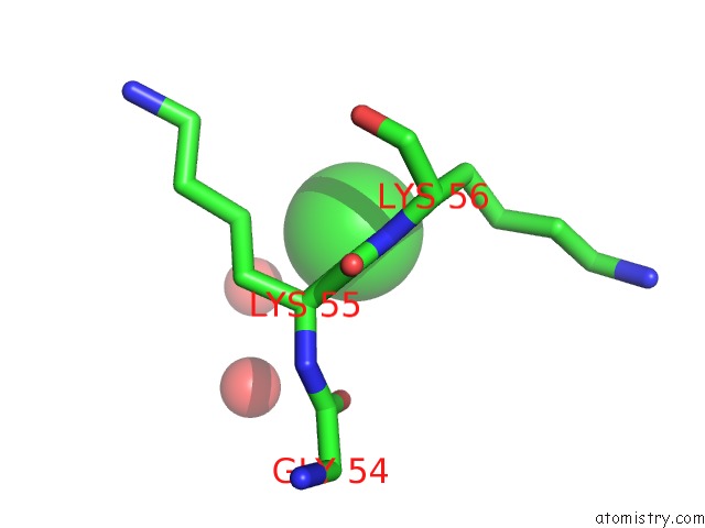
Mono view
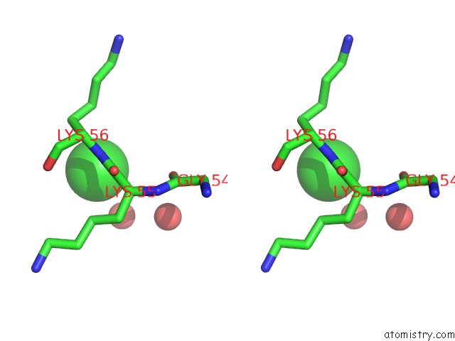
Stereo pair view
Mono view
Stereo pair view
A full contact list of Chlorine with other atoms in the Cl binding
site number 2 of Crystal Structure of the Hexameric Purine Nucleoside Phosphorylase From Bacillus Subtilis in Complex with Hypoxanthine within 5.0Å range:
|
Chlorine binding site 3 out of 3 in 4dab
Go back to
Chlorine binding site 3 out
of 3 in the Crystal Structure of the Hexameric Purine Nucleoside Phosphorylase From Bacillus Subtilis in Complex with Hypoxanthine
Mono view
Stereo pair view
Mono view
Stereo pair view
A full contact list of Chlorine with other atoms in the Cl binding
site number 3 of Crystal Structure of the Hexameric Purine Nucleoside Phosphorylase From Bacillus Subtilis in Complex with Hypoxanthine within 5.0Å range:
|
Reference:
P.O.De Giuseppe,
N.H.Martins,
A.N.Meza,
C.R.Dos Santos,
H.D.Pereira,
M.T.Murakami.
Insights Into Phosphate Cooperativity and Influence of Substrate Modifications on Binding and Catalysis of Hexameric Purine Nucleoside Phosphorylases. Plos One V. 7 44282 2012.
ISSN: ESSN 1932-6203
PubMed: 22957058
DOI: 10.1371/JOURNAL.PONE.0044282
Page generated: Sat Dec 12 10:31:37 2020
ISSN: ESSN 1932-6203
PubMed: 22957058
DOI: 10.1371/JOURNAL.PONE.0044282
Last articles
Zn in 8WB0Zn in 8WAX
Zn in 8WAU
Zn in 8WAZ
Zn in 8WAY
Zn in 8WAV
Zn in 8WAW
Zn in 8WAT
Zn in 8W7M
Zn in 8WD3