Chlorine in PDB 4ws6: Crystal Structure of Mycobacterium Tuberculosis Uracil-Dna Glycosylase in Complex with 5-Aminouracil, Form I
Enzymatic activity of Crystal Structure of Mycobacterium Tuberculosis Uracil-Dna Glycosylase in Complex with 5-Aminouracil, Form I
All present enzymatic activity of Crystal Structure of Mycobacterium Tuberculosis Uracil-Dna Glycosylase in Complex with 5-Aminouracil, Form I:
3.2.2.27;
3.2.2.27;
Protein crystallography data
The structure of Crystal Structure of Mycobacterium Tuberculosis Uracil-Dna Glycosylase in Complex with 5-Aminouracil, Form I, PDB code: 4ws6
was solved by
S.M.Arif,
K.Geethanandan,
P.Mishra,
A.Surolia,
U.Varshney,
M.Vijayan,
with X-Ray Crystallography technique. A brief refinement statistics is given in the table below:
| Resolution Low / High (Å) | 25.42 / 1.10 |
| Space group | P 1 21 1 |
| Cell size a, b, c (Å), α, β, γ (°) | 38.958, 63.976, 45.225, 90.00, 112.36, 90.00 |
| R / Rfree (%) | 12.8 / 14.6 |
Chlorine Binding Sites:
The binding sites of Chlorine atom in the Crystal Structure of Mycobacterium Tuberculosis Uracil-Dna Glycosylase in Complex with 5-Aminouracil, Form I
(pdb code 4ws6). This binding sites where shown within
5.0 Angstroms radius around Chlorine atom.
In total 2 binding sites of Chlorine where determined in the Crystal Structure of Mycobacterium Tuberculosis Uracil-Dna Glycosylase in Complex with 5-Aminouracil, Form I, PDB code: 4ws6:
Jump to Chlorine binding site number: 1; 2;
In total 2 binding sites of Chlorine where determined in the Crystal Structure of Mycobacterium Tuberculosis Uracil-Dna Glycosylase in Complex with 5-Aminouracil, Form I, PDB code: 4ws6:
Jump to Chlorine binding site number: 1; 2;
Chlorine binding site 1 out of 2 in 4ws6
Go back to
Chlorine binding site 1 out
of 2 in the Crystal Structure of Mycobacterium Tuberculosis Uracil-Dna Glycosylase in Complex with 5-Aminouracil, Form I
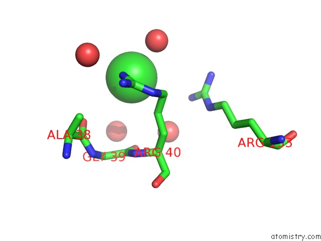
Mono view
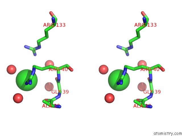
Stereo pair view
Mono view
Stereo pair view
A full contact list of Chlorine with other atoms in the Cl binding
site number 1 of Crystal Structure of Mycobacterium Tuberculosis Uracil-Dna Glycosylase in Complex with 5-Aminouracil, Form I within 5.0Å range:
|
Chlorine binding site 2 out of 2 in 4ws6
Go back to
Chlorine binding site 2 out
of 2 in the Crystal Structure of Mycobacterium Tuberculosis Uracil-Dna Glycosylase in Complex with 5-Aminouracil, Form I
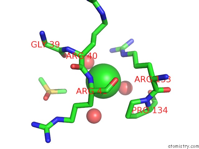
Mono view
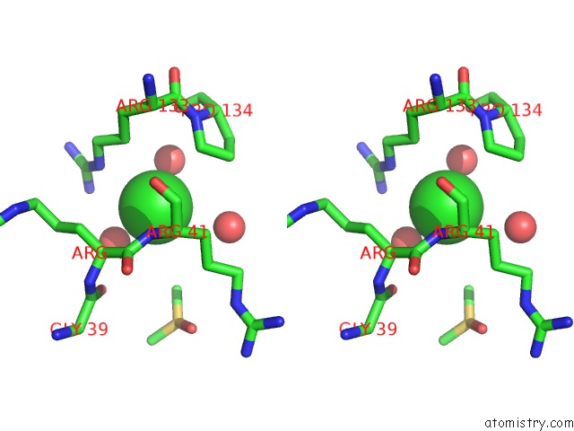
Stereo pair view
Mono view
Stereo pair view
A full contact list of Chlorine with other atoms in the Cl binding
site number 2 of Crystal Structure of Mycobacterium Tuberculosis Uracil-Dna Glycosylase in Complex with 5-Aminouracil, Form I within 5.0Å range:
|
Reference:
S.M.Arif,
K.Geethanandan,
P.Mishra,
A.Surolia,
U.Varshney,
M.Vijayan.
Structural Plasticity in Mycobacterium Tuberculosis Uracil-Dna Glycosylase (Mtung) and Its Functional Implications. Acta Crystallogr.,Sect.D V. 71 1514 2015.
ISSN: ESSN 1399-0047
PubMed: 26143923
DOI: 10.1107/S1399004715009311
Page generated: Fri Jul 26 03:04:45 2024
ISSN: ESSN 1399-0047
PubMed: 26143923
DOI: 10.1107/S1399004715009311
Last articles
Zn in 9J0NZn in 9J0O
Zn in 9J0P
Zn in 9FJX
Zn in 9EKB
Zn in 9C0F
Zn in 9CAH
Zn in 9CH0
Zn in 9CH3
Zn in 9CH1