Chlorine in PDB 4yvf: Structure of S-Adenosyl-L-Homocysteine Hydrolase
Enzymatic activity of Structure of S-Adenosyl-L-Homocysteine Hydrolase
All present enzymatic activity of Structure of S-Adenosyl-L-Homocysteine Hydrolase:
3.3.1.1;
3.3.1.1;
Protein crystallography data
The structure of Structure of S-Adenosyl-L-Homocysteine Hydrolase, PDB code: 4yvf
was solved by
K.Akiko,
with X-Ray Crystallography technique. A brief refinement statistics is given in the table below:
| Resolution Low / High (Å) | 47.81 / 2.70 |
| Space group | C 2 2 21 |
| Cell size a, b, c (Å), α, β, γ (°) | 91.528, 134.150, 185.164, 90.00, 90.00, 90.00 |
| R / Rfree (%) | n/a / n/a |
Chlorine Binding Sites:
The binding sites of Chlorine atom in the Structure of S-Adenosyl-L-Homocysteine Hydrolase
(pdb code 4yvf). This binding sites where shown within
5.0 Angstroms radius around Chlorine atom.
In total 4 binding sites of Chlorine where determined in the Structure of S-Adenosyl-L-Homocysteine Hydrolase, PDB code: 4yvf:
Jump to Chlorine binding site number: 1; 2; 3; 4;
In total 4 binding sites of Chlorine where determined in the Structure of S-Adenosyl-L-Homocysteine Hydrolase, PDB code: 4yvf:
Jump to Chlorine binding site number: 1; 2; 3; 4;
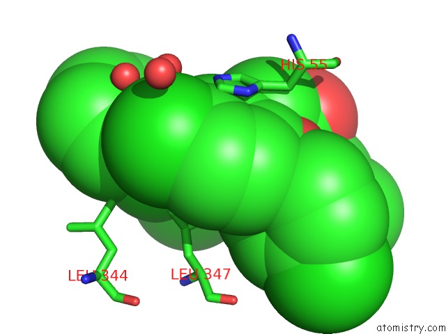
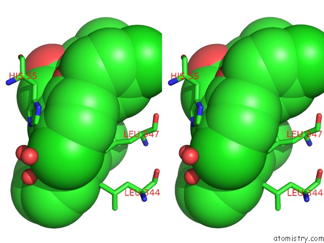
Chlorine binding site 1 out of 4 in 4yvf
Go back to
Chlorine binding site 1 out
of 4 in the Structure of S-Adenosyl-L-Homocysteine Hydrolase
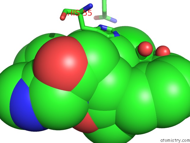
Mono view
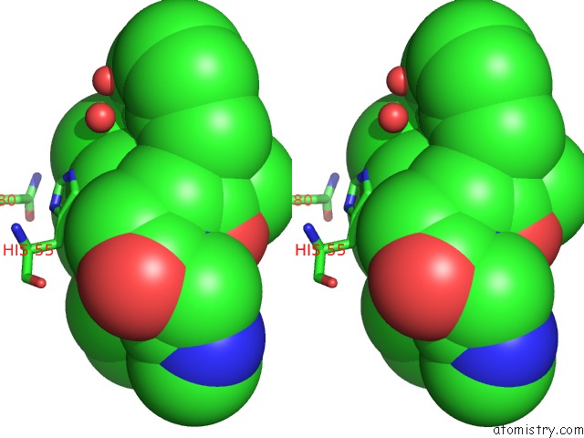
Stereo pair view
Mono view
Stereo pair view
A full contact list of Chlorine with other atoms in the Cl binding
site number 1 of Structure of S-Adenosyl-L-Homocysteine Hydrolase within 5.0Å range:
|
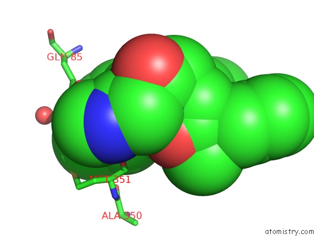
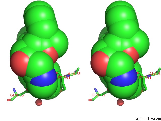
Chlorine binding site 2 out of 4 in 4yvf
Go back to
Chlorine binding site 2 out
of 4 in the Structure of S-Adenosyl-L-Homocysteine Hydrolase
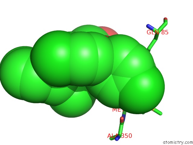
Mono view
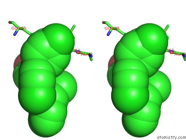
Stereo pair view
Mono view
Stereo pair view
A full contact list of Chlorine with other atoms in the Cl binding
site number 2 of Structure of S-Adenosyl-L-Homocysteine Hydrolase within 5.0Å range:
|
Chlorine binding site 3 out of 4 in 4yvf
Go back to
Chlorine binding site 3 out
of 4 in the Structure of S-Adenosyl-L-Homocysteine Hydrolase
Mono view
Stereo pair view
Mono view
Stereo pair view
A full contact list of Chlorine with other atoms in the Cl binding
site number 3 of Structure of S-Adenosyl-L-Homocysteine Hydrolase within 5.0Å range:
|
Chlorine binding site 4 out of 4 in 4yvf
Go back to
Chlorine binding site 4 out
of 4 in the Structure of S-Adenosyl-L-Homocysteine Hydrolase
Mono view
Stereo pair view
Mono view
Stereo pair view
A full contact list of Chlorine with other atoms in the Cl binding
site number 4 of Structure of S-Adenosyl-L-Homocysteine Hydrolase within 5.0Å range:
|
Reference:
A.Nakao,
H.Suzuki,
H.Ueno,
H.Iwasaki,
T.Setsuta,
A.Kashima,
S.Sunada.
Discovery and Structural Analyses of S-Adenosyl-L-Homocysteine Hydrolase Inhibitors Based on Non-Adenosine Analogs. Bioorg.Med.Chem. V. 23 4952 2015.
ISSN: ESSN 1464-3391
PubMed: 26037610
DOI: 10.1016/J.BMC.2015.05.018
Page generated: Sat Dec 12 11:26:28 2020
ISSN: ESSN 1464-3391
PubMed: 26037610
DOI: 10.1016/J.BMC.2015.05.018
Last articles
Zn in 8WB0Zn in 8WAX
Zn in 8WAU
Zn in 8WAZ
Zn in 8WAY
Zn in 8WAV
Zn in 8WAW
Zn in 8WAT
Zn in 8W7M
Zn in 8WD3