Chlorine in PDB 7b1n: Crystal Structure of Phosphatidyl Serine Synthase (Pss) in the Closed Conformation with Bound Citrate.
Enzymatic activity of Crystal Structure of Phosphatidyl Serine Synthase (Pss) in the Closed Conformation with Bound Citrate.
All present enzymatic activity of Crystal Structure of Phosphatidyl Serine Synthase (Pss) in the Closed Conformation with Bound Citrate.:
2.7.8.8;
2.7.8.8;
Protein crystallography data
The structure of Crystal Structure of Phosphatidyl Serine Synthase (Pss) in the Closed Conformation with Bound Citrate., PDB code: 7b1n
was solved by
O.Yildiz,
M.Centola,
with X-Ray Crystallography technique. A brief refinement statistics is given in the table below:
| Resolution Low / High (Å) | 46.70 / 2.80 |
| Space group | P 2 21 21 |
| Cell size a, b, c (Å), α, β, γ (°) | 62.13, 70.81, 94.09, 90, 90, 90 |
| R / Rfree (%) | 30.2 / 35.5 |
Other elements in 7b1n:
The structure of Crystal Structure of Phosphatidyl Serine Synthase (Pss) in the Closed Conformation with Bound Citrate. also contains other interesting chemical elements:
| Calcium | (Ca) | 2 atoms |
Chlorine Binding Sites:
The binding sites of Chlorine atom in the Crystal Structure of Phosphatidyl Serine Synthase (Pss) in the Closed Conformation with Bound Citrate.
(pdb code 7b1n). This binding sites where shown within
5.0 Angstroms radius around Chlorine atom.
In total 4 binding sites of Chlorine where determined in the Crystal Structure of Phosphatidyl Serine Synthase (Pss) in the Closed Conformation with Bound Citrate., PDB code: 7b1n:
Jump to Chlorine binding site number: 1; 2; 3; 4;
In total 4 binding sites of Chlorine where determined in the Crystal Structure of Phosphatidyl Serine Synthase (Pss) in the Closed Conformation with Bound Citrate., PDB code: 7b1n:
Jump to Chlorine binding site number: 1; 2; 3; 4;
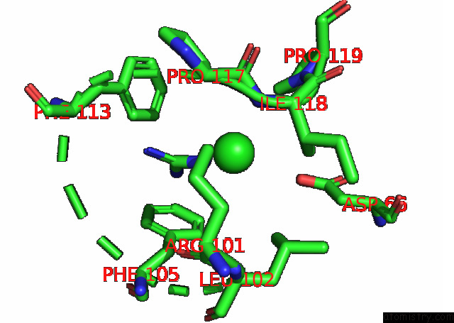
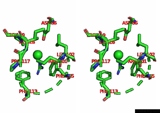
Chlorine binding site 1 out of 4 in 7b1n
Go back to
Chlorine binding site 1 out
of 4 in the Crystal Structure of Phosphatidyl Serine Synthase (Pss) in the Closed Conformation with Bound Citrate.
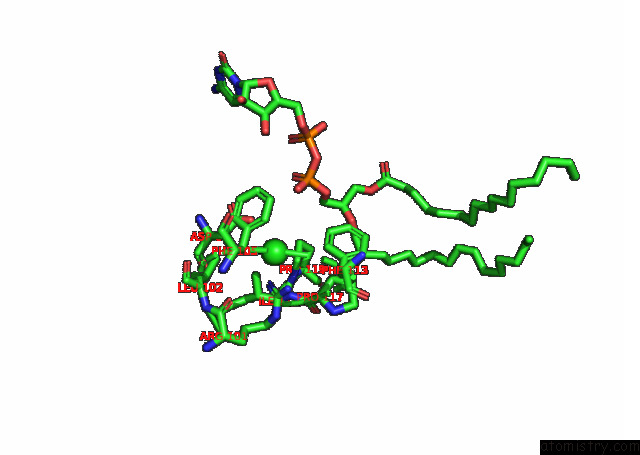
Mono view
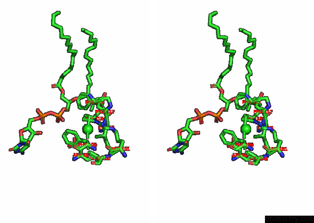
Stereo pair view
Mono view
Stereo pair view
A full contact list of Chlorine with other atoms in the Cl binding
site number 1 of Crystal Structure of Phosphatidyl Serine Synthase (Pss) in the Closed Conformation with Bound Citrate. within 5.0Å range:
|
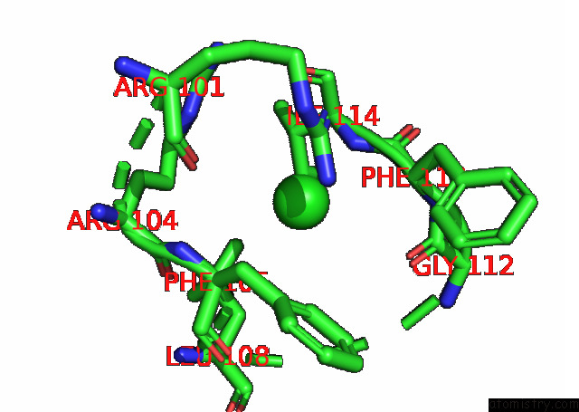
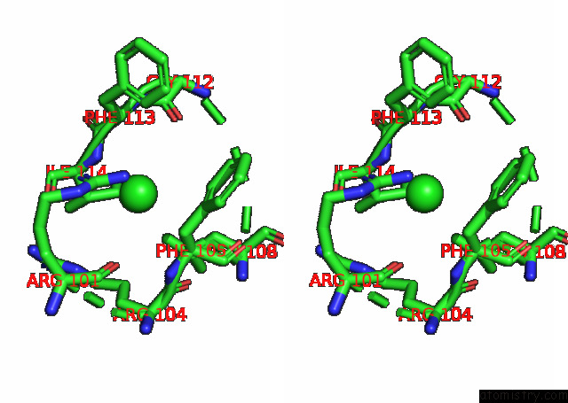
Chlorine binding site 2 out of 4 in 7b1n
Go back to
Chlorine binding site 2 out
of 4 in the Crystal Structure of Phosphatidyl Serine Synthase (Pss) in the Closed Conformation with Bound Citrate.
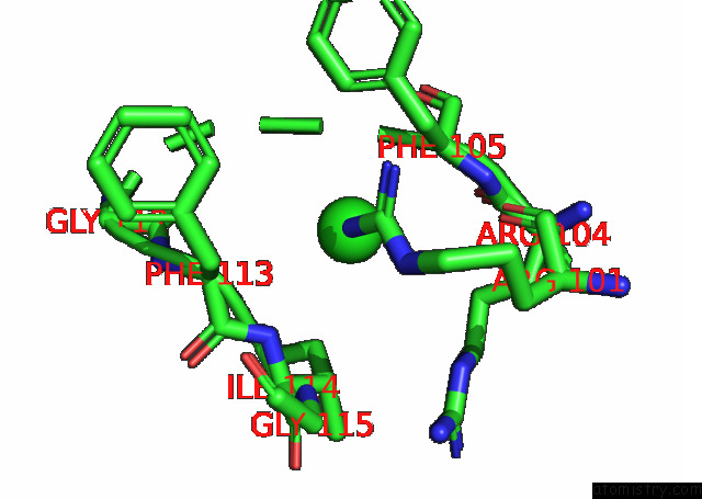
Mono view
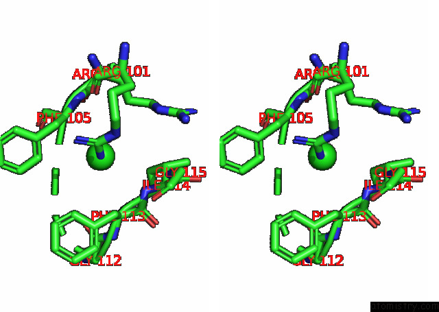
Stereo pair view
Mono view
Stereo pair view
A full contact list of Chlorine with other atoms in the Cl binding
site number 2 of Crystal Structure of Phosphatidyl Serine Synthase (Pss) in the Closed Conformation with Bound Citrate. within 5.0Å range:
|
Chlorine binding site 3 out of 4 in 7b1n
Go back to
Chlorine binding site 3 out
of 4 in the Crystal Structure of Phosphatidyl Serine Synthase (Pss) in the Closed Conformation with Bound Citrate.
Mono view
Stereo pair view
Mono view
Stereo pair view
A full contact list of Chlorine with other atoms in the Cl binding
site number 3 of Crystal Structure of Phosphatidyl Serine Synthase (Pss) in the Closed Conformation with Bound Citrate. within 5.0Å range:
|
Chlorine binding site 4 out of 4 in 7b1n
Go back to
Chlorine binding site 4 out
of 4 in the Crystal Structure of Phosphatidyl Serine Synthase (Pss) in the Closed Conformation with Bound Citrate.
Mono view
Stereo pair view
Mono view
Stereo pair view
A full contact list of Chlorine with other atoms in the Cl binding
site number 4 of Crystal Structure of Phosphatidyl Serine Synthase (Pss) in the Closed Conformation with Bound Citrate. within 5.0Å range:
|
Reference:
M.Centola,
H.Betz,
O.Yildiz.
Crystal Structures of Phosphatidyl Serine Synthase Pss Reveal the Catalytic Mechanism of Cdp-Dag Alcohol O-Phosphatidyl Transferases Nat Commun V. 12 6982 2021.
ISSN: ESSN 2041-1723
DOI: 10.1038/S41467-021-27281-W
Page generated: Mon Jul 29 18:57:53 2024
ISSN: ESSN 2041-1723
DOI: 10.1038/S41467-021-27281-W
Last articles
Zn in 9J0NZn in 9J0O
Zn in 9J0P
Zn in 9FJX
Zn in 9EKB
Zn in 9C0F
Zn in 9CAH
Zn in 9CH0
Zn in 9CH3
Zn in 9CH1