Chlorine in PDB 7jhi: Structure of Human Beta 1,3-N-Acetylglucosaminyltransferase 2 Iodide- Derivative
Enzymatic activity of Structure of Human Beta 1,3-N-Acetylglucosaminyltransferase 2 Iodide- Derivative
All present enzymatic activity of Structure of Human Beta 1,3-N-Acetylglucosaminyltransferase 2 Iodide- Derivative:
2.4.1.149;
2.4.1.149;
Protein crystallography data
The structure of Structure of Human Beta 1,3-N-Acetylglucosaminyltransferase 2 Iodide- Derivative, PDB code: 7jhi
was solved by
Y.Hao,
X.Huang,
with X-Ray Crystallography technique. A brief refinement statistics is given in the table below:
| Resolution Low / High (Å) | 47.33 / 2.50 |
| Space group | P 1 21 1 |
| Cell size a, b, c (Å), α, β, γ (°) | 67.431, 81.385, 157.138, 90.00, 98.63, 90.00 |
| R / Rfree (%) | 20.2 / 24.8 |
Other elements in 7jhi:
The structure of Structure of Human Beta 1,3-N-Acetylglucosaminyltransferase 2 Iodide- Derivative also contains other interesting chemical elements:
| Magnesium | (Mg) | 3 atoms |
| Iodine | (I) | 41 atoms |
Chlorine Binding Sites:
The binding sites of Chlorine atom in the Structure of Human Beta 1,3-N-Acetylglucosaminyltransferase 2 Iodide- Derivative
(pdb code 7jhi). This binding sites where shown within
5.0 Angstroms radius around Chlorine atom.
In total 5 binding sites of Chlorine where determined in the Structure of Human Beta 1,3-N-Acetylglucosaminyltransferase 2 Iodide- Derivative, PDB code: 7jhi:
Jump to Chlorine binding site number: 1; 2; 3; 4; 5;
In total 5 binding sites of Chlorine where determined in the Structure of Human Beta 1,3-N-Acetylglucosaminyltransferase 2 Iodide- Derivative, PDB code: 7jhi:
Jump to Chlorine binding site number: 1; 2; 3; 4; 5;
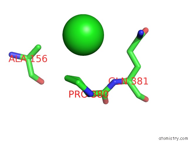
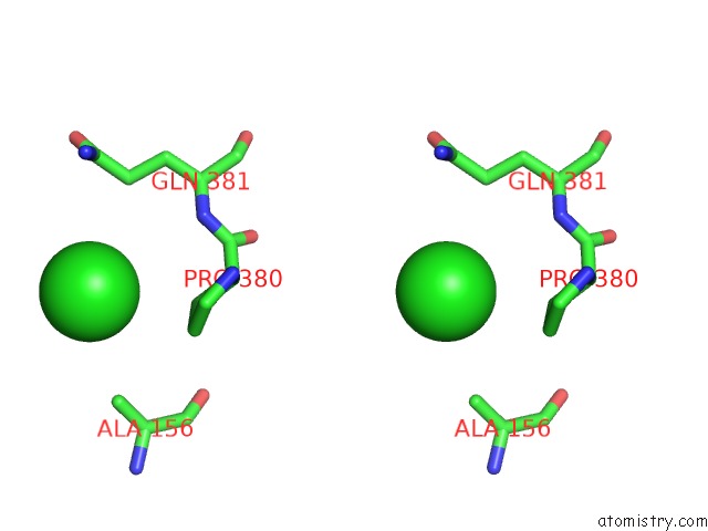
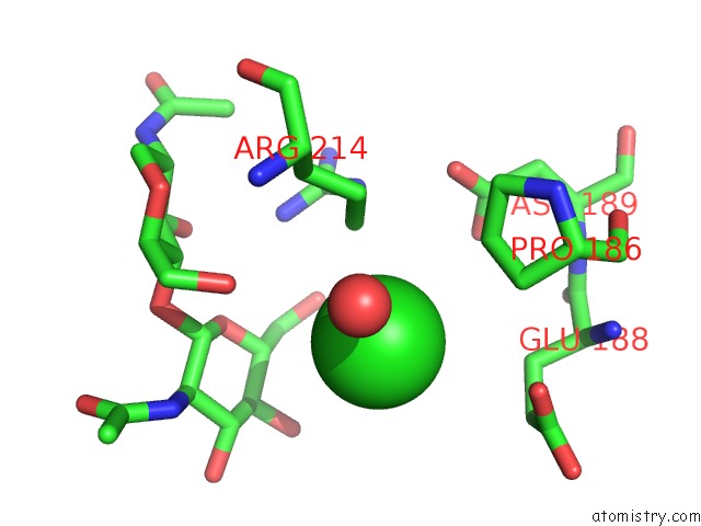
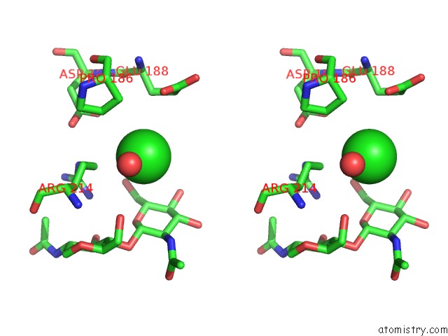
Chlorine binding site 1 out of 5 in 7jhi
Go back to
Chlorine binding site 1 out
of 5 in the Structure of Human Beta 1,3-N-Acetylglucosaminyltransferase 2 Iodide- Derivative
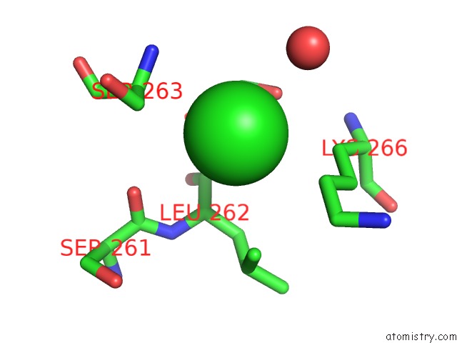
Mono view
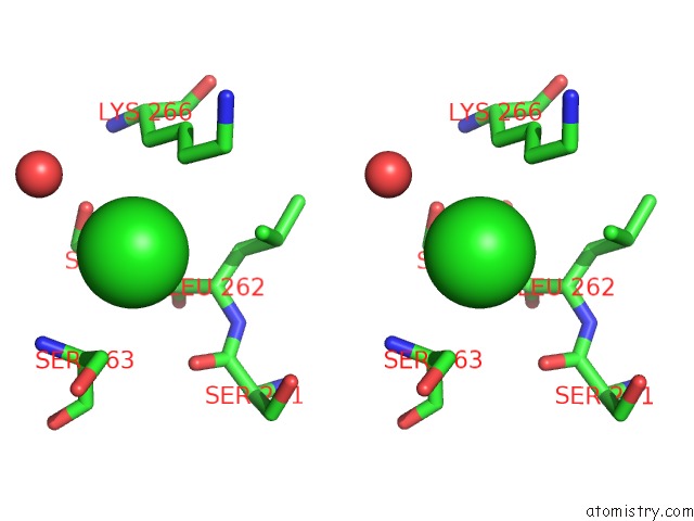
Stereo pair view
Mono view
Stereo pair view
A full contact list of Chlorine with other atoms in the Cl binding
site number 1 of Structure of Human Beta 1,3-N-Acetylglucosaminyltransferase 2 Iodide- Derivative within 5.0Å range:
|
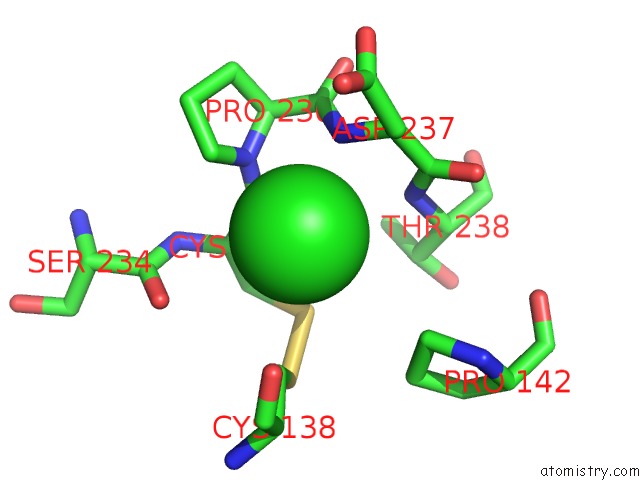
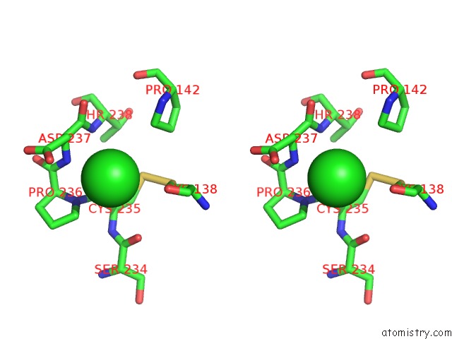
Chlorine binding site 2 out of 5 in 7jhi
Go back to
Chlorine binding site 2 out
of 5 in the Structure of Human Beta 1,3-N-Acetylglucosaminyltransferase 2 Iodide- Derivative
Mono view
Stereo pair view
Mono view
Stereo pair view
A full contact list of Chlorine with other atoms in the Cl binding
site number 2 of Structure of Human Beta 1,3-N-Acetylglucosaminyltransferase 2 Iodide- Derivative within 5.0Å range:
|
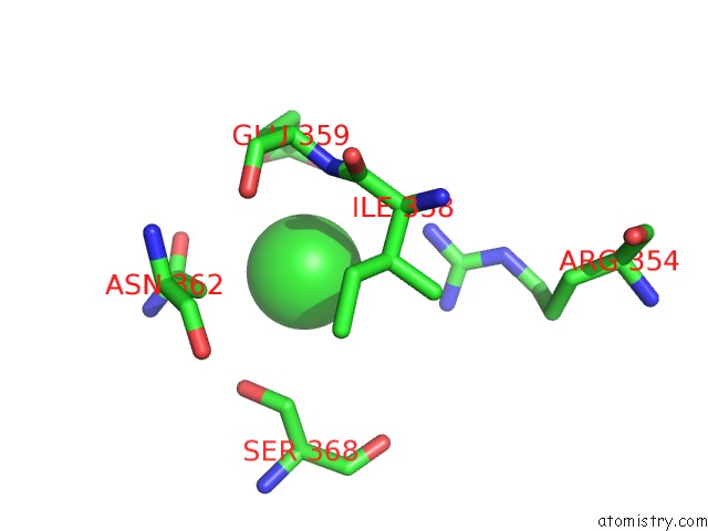
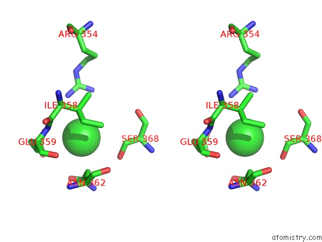
Chlorine binding site 3 out of 5 in 7jhi
Go back to
Chlorine binding site 3 out
of 5 in the Structure of Human Beta 1,3-N-Acetylglucosaminyltransferase 2 Iodide- Derivative
Mono view
Stereo pair view
Mono view
Stereo pair view
A full contact list of Chlorine with other atoms in the Cl binding
site number 3 of Structure of Human Beta 1,3-N-Acetylglucosaminyltransferase 2 Iodide- Derivative within 5.0Å range:
|
Chlorine binding site 4 out of 5 in 7jhi
Go back to
Chlorine binding site 4 out
of 5 in the Structure of Human Beta 1,3-N-Acetylglucosaminyltransferase 2 Iodide- Derivative
Mono view
Stereo pair view
Mono view
Stereo pair view
A full contact list of Chlorine with other atoms in the Cl binding
site number 4 of Structure of Human Beta 1,3-N-Acetylglucosaminyltransferase 2 Iodide- Derivative within 5.0Å range:
|
Chlorine binding site 5 out of 5 in 7jhi
Go back to
Chlorine binding site 5 out
of 5 in the Structure of Human Beta 1,3-N-Acetylglucosaminyltransferase 2 Iodide- Derivative
Mono view
Stereo pair view
Mono view
Stereo pair view
A full contact list of Chlorine with other atoms in the Cl binding
site number 5 of Structure of Human Beta 1,3-N-Acetylglucosaminyltransferase 2 Iodide- Derivative within 5.0Å range:
|
Reference:
Y.Hao,
A.Crequer-Grandhomme,
N.Javier,
A.Singh,
H.Chen,
P.Manzanillo,
M.C.Lo,
X.Huang.
Structures and Mechanism of Human Glycosyltransferase Beta 1,3-N-Acetylglucosaminyltransferase 2 (B3GNT2), An Important Player in Immune Homeostasis. J.Biol.Chem. 2020.
ISSN: ESSN 1083-351X
PubMed: 33158990
DOI: 10.1074/JBC.RA120.015306
Page generated: Mon Jul 29 23:04:33 2024
ISSN: ESSN 1083-351X
PubMed: 33158990
DOI: 10.1074/JBC.RA120.015306
Last articles
Zn in 9J0NZn in 9J0O
Zn in 9J0P
Zn in 9FJX
Zn in 9EKB
Zn in 9C0F
Zn in 9CAH
Zn in 9CH0
Zn in 9CH3
Zn in 9CH1