Chlorine in PDB 7jw4: Crystal Structure of PDGH110B in Complex with D-Galactose
Protein crystallography data
The structure of Crystal Structure of PDGH110B in Complex with D-Galactose, PDB code: 7jw4
was solved by
A.G.Hettle,
A.B.Boraston,
with X-Ray Crystallography technique. A brief refinement statistics is given in the table below:
| Resolution Low / High (Å) | 24.98 / 2.34 |
| Space group | C 1 2 1 |
| Cell size a, b, c (Å), α, β, γ (°) | 169.786, 128.093, 100.014, 90.00, 123.16, 90.00 |
| R / Rfree (%) | 18.5 / 23.1 |
Other elements in 7jw4:
The structure of Crystal Structure of PDGH110B in Complex with D-Galactose also contains other interesting chemical elements:
| Nickel | (Ni) | 2 atoms |
Chlorine Binding Sites:
The binding sites of Chlorine atom in the Crystal Structure of PDGH110B in Complex with D-Galactose
(pdb code 7jw4). This binding sites where shown within
5.0 Angstroms radius around Chlorine atom.
In total 2 binding sites of Chlorine where determined in the Crystal Structure of PDGH110B in Complex with D-Galactose, PDB code: 7jw4:
Jump to Chlorine binding site number: 1; 2;
In total 2 binding sites of Chlorine where determined in the Crystal Structure of PDGH110B in Complex with D-Galactose, PDB code: 7jw4:
Jump to Chlorine binding site number: 1; 2;
Chlorine binding site 1 out of 2 in 7jw4
Go back to
Chlorine binding site 1 out
of 2 in the Crystal Structure of PDGH110B in Complex with D-Galactose
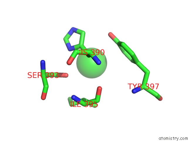
Mono view
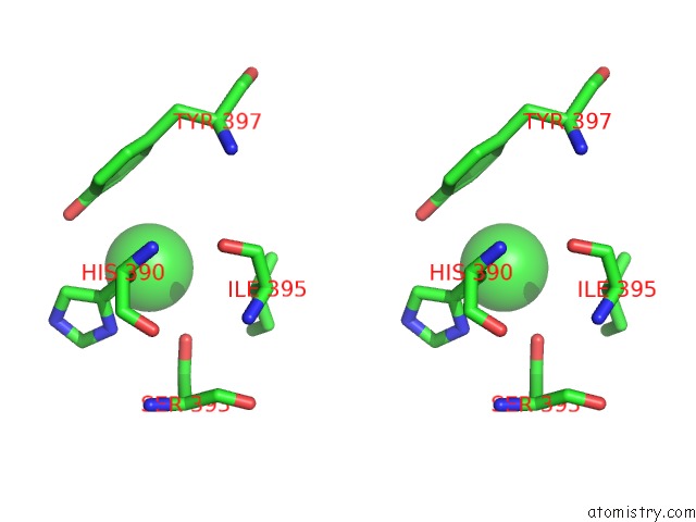
Stereo pair view
Mono view
Stereo pair view
A full contact list of Chlorine with other atoms in the Cl binding
site number 1 of Crystal Structure of PDGH110B in Complex with D-Galactose within 5.0Å range:
|
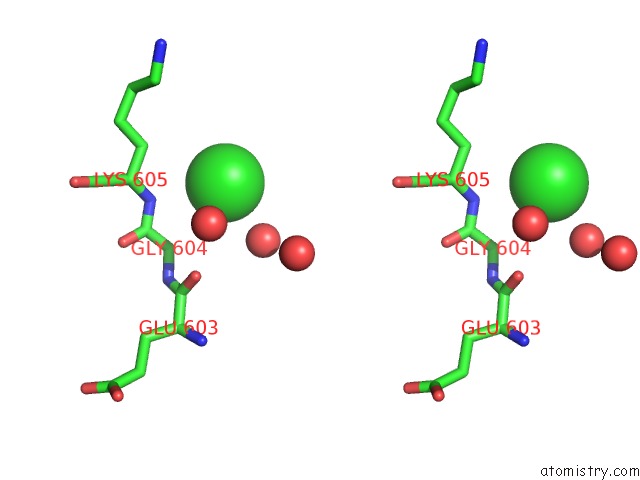
Chlorine binding site 2 out of 2 in 7jw4
Go back to
Chlorine binding site 2 out
of 2 in the Crystal Structure of PDGH110B in Complex with D-Galactose
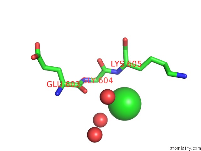
Mono view
Stereo pair view
Mono view
Stereo pair view
A full contact list of Chlorine with other atoms in the Cl binding
site number 2 of Crystal Structure of PDGH110B in Complex with D-Galactose within 5.0Å range:
|
Reference:
B.E.Mcguire,
A.Hettle,
C.Vickers,
D.T.King,
D.J.Vocadlo,
A.B.Boraston.
The Structure of A Family 110 Glycoside Hydrolase Provides Insight Into the Hydrolysis of Alpha-(1,3)-Galactosidic Linkages in Lambda-Carrageenan and Blood Group Antigens. J.Biol.Chem. 2020.
ISSN: ESSN 1083-351X
PubMed: 33127644
DOI: 10.1074/JBC.RA120.015776
Page generated: Mon Jul 29 23:17:44 2024
ISSN: ESSN 1083-351X
PubMed: 33127644
DOI: 10.1074/JBC.RA120.015776
Last articles
Zn in 9J0NZn in 9J0O
Zn in 9J0P
Zn in 9FJX
Zn in 9EKB
Zn in 9C0F
Zn in 9CAH
Zn in 9CH0
Zn in 9CH3
Zn in 9CH1