Chlorine in PDB 7k31: Crystal Structure of Endonuclease Q Complex with 27-Mer Duplex Substrate with Di at the Active Site
Protein crystallography data
The structure of Crystal Structure of Endonuclease Q Complex with 27-Mer Duplex Substrate with Di at the Active Site, PDB code: 7k31
was solved by
K.Shi,
N.M.Moeller,
S.Banerjee,
L.Yin,
K.Orellana,
H.Aihara,
with X-Ray Crystallography technique. A brief refinement statistics is given in the table below:
| Resolution Low / High (Å) | 87.47 / 2.88 |
| Space group | H 3 |
| Cell size a, b, c (Å), α, β, γ (°) | 151.12, 151.12, 117.58, 90, 90, 120 |
| R / Rfree (%) | 18.2 / 23.8 |
Other elements in 7k31:
The structure of Crystal Structure of Endonuclease Q Complex with 27-Mer Duplex Substrate with Di at the Active Site also contains other interesting chemical elements:
| Magnesium | (Mg) | 1 atom |
| Zinc | (Zn) | 3 atoms |
Chlorine Binding Sites:
The binding sites of Chlorine atom in the Crystal Structure of Endonuclease Q Complex with 27-Mer Duplex Substrate with Di at the Active Site
(pdb code 7k31). This binding sites where shown within
5.0 Angstroms radius around Chlorine atom.
In total 2 binding sites of Chlorine where determined in the Crystal Structure of Endonuclease Q Complex with 27-Mer Duplex Substrate with Di at the Active Site, PDB code: 7k31:
Jump to Chlorine binding site number: 1; 2;
In total 2 binding sites of Chlorine where determined in the Crystal Structure of Endonuclease Q Complex with 27-Mer Duplex Substrate with Di at the Active Site, PDB code: 7k31:
Jump to Chlorine binding site number: 1; 2;
Chlorine binding site 1 out of 2 in 7k31
Go back to
Chlorine binding site 1 out
of 2 in the Crystal Structure of Endonuclease Q Complex with 27-Mer Duplex Substrate with Di at the Active Site
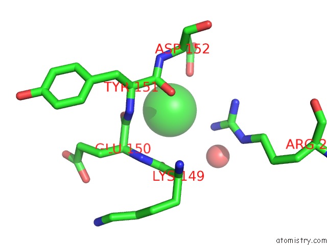
Mono view
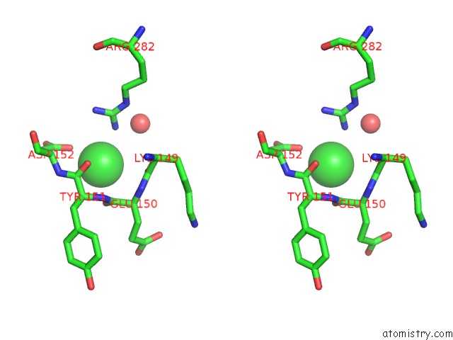
Stereo pair view
Mono view
Stereo pair view
A full contact list of Chlorine with other atoms in the Cl binding
site number 1 of Crystal Structure of Endonuclease Q Complex with 27-Mer Duplex Substrate with Di at the Active Site within 5.0Å range:
|
Chlorine binding site 2 out of 2 in 7k31
Go back to
Chlorine binding site 2 out
of 2 in the Crystal Structure of Endonuclease Q Complex with 27-Mer Duplex Substrate with Di at the Active Site
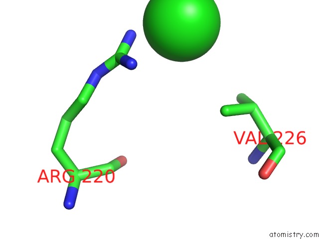
Mono view
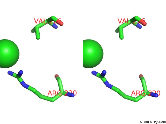
Stereo pair view
Mono view
Stereo pair view
A full contact list of Chlorine with other atoms in the Cl binding
site number 2 of Crystal Structure of Endonuclease Q Complex with 27-Mer Duplex Substrate with Di at the Active Site within 5.0Å range:
|
Reference:
K.Shi,
N.H.Moeller,
S.Banerjee,
J.L.Mccann,
M.A.Carpenter,
L.Yin,
R.Moorthy,
K.Orellana,
D.A.Harki,
R.S.Harris,
H.Aihara.
Structural Basis For Recognition of Distinct Deaminated Dna Lesions By Endonuclease Q. Proc.Natl.Acad.Sci.Usa V. 118 2021.
ISSN: ESSN 1091-6490
PubMed: 33658373
DOI: 10.1073/PNAS.2021120118
Page generated: Mon Jul 29 23:21:03 2024
ISSN: ESSN 1091-6490
PubMed: 33658373
DOI: 10.1073/PNAS.2021120118
Last articles
Zn in 9J0NZn in 9J0O
Zn in 9J0P
Zn in 9FJX
Zn in 9EKB
Zn in 9C0F
Zn in 9CAH
Zn in 9CH0
Zn in 9CH3
Zn in 9CH1