Chlorine in PDB 7kdr: Crystal Structure of Escherichia Coli Hppk in Complex with Bisubstrate Inhibitor Hp-75
Enzymatic activity of Crystal Structure of Escherichia Coli Hppk in Complex with Bisubstrate Inhibitor Hp-75
All present enzymatic activity of Crystal Structure of Escherichia Coli Hppk in Complex with Bisubstrate Inhibitor Hp-75:
2.7.6.3;
2.7.6.3;
Protein crystallography data
The structure of Crystal Structure of Escherichia Coli Hppk in Complex with Bisubstrate Inhibitor Hp-75, PDB code: 7kdr
was solved by
G.X.Shaw,
G.Shi,
X.Ji,
with X-Ray Crystallography technique. A brief refinement statistics is given in the table below:
| Resolution Low / High (Å) | 28.97 / 1.49 |
| Space group | C 1 2 1 |
| Cell size a, b, c (Å), α, β, γ (°) | 61.446, 42.797, 56.283, 90.00, 101.54, 90.00 |
| R / Rfree (%) | 16.2 / 20.1 |
Chlorine Binding Sites:
The binding sites of Chlorine atom in the Crystal Structure of Escherichia Coli Hppk in Complex with Bisubstrate Inhibitor Hp-75
(pdb code 7kdr). This binding sites where shown within
5.0 Angstroms radius around Chlorine atom.
In total only one binding site of Chlorine was determined in the Crystal Structure of Escherichia Coli Hppk in Complex with Bisubstrate Inhibitor Hp-75, PDB code: 7kdr:
In total only one binding site of Chlorine was determined in the Crystal Structure of Escherichia Coli Hppk in Complex with Bisubstrate Inhibitor Hp-75, PDB code: 7kdr:
Chlorine binding site 1 out of 1 in 7kdr
Go back to
Chlorine binding site 1 out
of 1 in the Crystal Structure of Escherichia Coli Hppk in Complex with Bisubstrate Inhibitor Hp-75
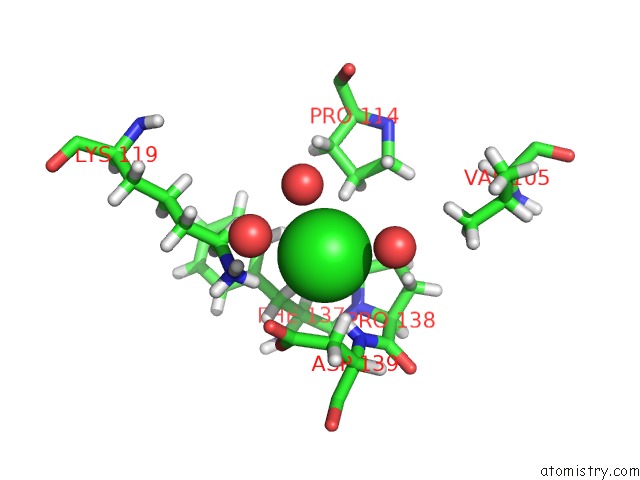
Mono view
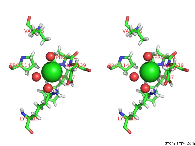
Stereo pair view
Mono view
Stereo pair view
A full contact list of Chlorine with other atoms in the Cl binding
site number 1 of Crystal Structure of Escherichia Coli Hppk in Complex with Bisubstrate Inhibitor Hp-75 within 5.0Å range:
|
Reference:
G.Shi,
G.X.Shaw,
F.Zhu,
S.G.Tarasov,
X.Ji.
Bisubstrate Inhibitors of 6-Hydroxymethyl-7,8-Dihydropterin Pyrophosphokinase: Transition State Analogs For High Affinity Binding. Bioorg.Med.Chem. 15847 2020.
ISSN: ESSN 1464-3391
PubMed: 33199204
DOI: 10.1016/J.BMC.2020.115847
Page generated: Mon Jul 29 23:30:49 2024
ISSN: ESSN 1464-3391
PubMed: 33199204
DOI: 10.1016/J.BMC.2020.115847
Last articles
Zn in 9JYWZn in 9IR4
Zn in 9IR3
Zn in 9GMX
Zn in 9GMW
Zn in 9JEJ
Zn in 9ERF
Zn in 9ERE
Zn in 9EGV
Zn in 9EGW