Chlorine in PDB 7lxg: Homocitrullinated Beta-Lactamase Oxa-48
Enzymatic activity of Homocitrullinated Beta-Lactamase Oxa-48
All present enzymatic activity of Homocitrullinated Beta-Lactamase Oxa-48:
3.5.2.6;
3.5.2.6;
Protein crystallography data
The structure of Homocitrullinated Beta-Lactamase Oxa-48, PDB code: 7lxg
was solved by
J.E.Serrano-Negron,
D.T.King,
D.J.Vocadlo,
with X-Ray Crystallography technique. A brief refinement statistics is given in the table below:
| Resolution Low / High (Å) | 47.12 / 2.20 |
| Space group | P 62 |
| Cell size a, b, c (Å), α, β, γ (°) | 143.96, 143.96, 56.03, 90, 90, 120 |
| R / Rfree (%) | 16.8 / 21.7 |
Chlorine Binding Sites:
The binding sites of Chlorine atom in the Homocitrullinated Beta-Lactamase Oxa-48
(pdb code 7lxg). This binding sites where shown within
5.0 Angstroms radius around Chlorine atom.
In total only one binding site of Chlorine was determined in the Homocitrullinated Beta-Lactamase Oxa-48, PDB code: 7lxg:
In total only one binding site of Chlorine was determined in the Homocitrullinated Beta-Lactamase Oxa-48, PDB code: 7lxg:
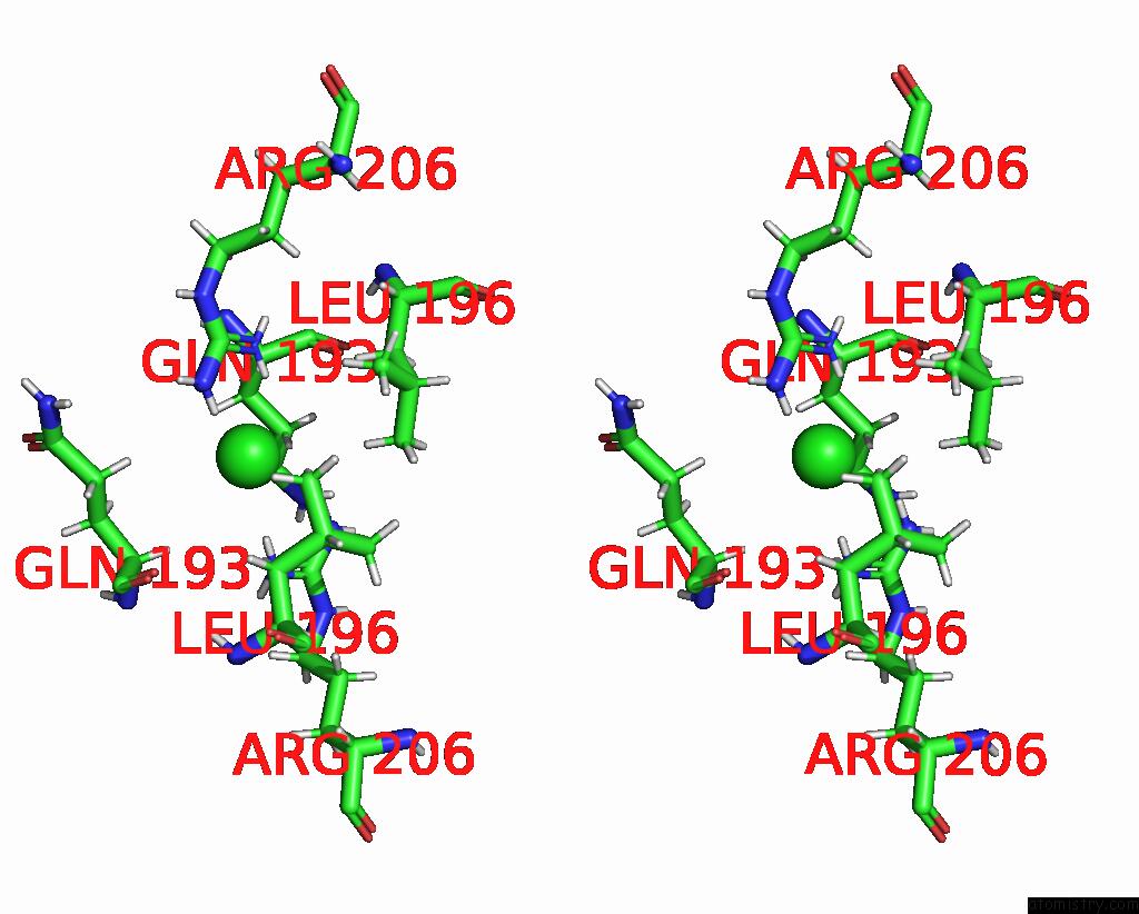
Chlorine binding site 1 out of 1 in 7lxg
Go back to
Chlorine binding site 1 out
of 1 in the Homocitrullinated Beta-Lactamase Oxa-48

Mono view
Stereo pair view
Mono view
Stereo pair view
A full contact list of Chlorine with other atoms in the Cl binding
site number 1 of Homocitrullinated Beta-Lactamase Oxa-48 within 5.0Å range:
|
Reference:
D.T.King,
S.Zhu,
D.B.Hardie,
J.E.Serrano-Negron,
Z.Madden,
S.Kolappan,
D.J.Vocadlo.
Chemoproteomic Identification of Co 2 -Dependent Lysine Carboxylation in Proteins. Nat.Chem.Biol. V. 18 782 2022.
ISSN: ESSN 1552-4469
PubMed: 35710617
DOI: 10.1038/S41589-022-01043-1
Page generated: Tue Jul 30 00:16:09 2024
ISSN: ESSN 1552-4469
PubMed: 35710617
DOI: 10.1038/S41589-022-01043-1
Last articles
Zn in 9JYWZn in 9IR4
Zn in 9IR3
Zn in 9GMX
Zn in 9GMW
Zn in 9JEJ
Zn in 9ERF
Zn in 9ERE
Zn in 9EGV
Zn in 9EGW