Chlorine in PDB 7mlt: Crystal Structure of Ricin A Chain in Complex with 5-(2-Ethylphenyl) Thiophene-2-Carboxylic Acid
Enzymatic activity of Crystal Structure of Ricin A Chain in Complex with 5-(2-Ethylphenyl) Thiophene-2-Carboxylic Acid
All present enzymatic activity of Crystal Structure of Ricin A Chain in Complex with 5-(2-Ethylphenyl) Thiophene-2-Carboxylic Acid:
3.2.2.22;
3.2.2.22;
Protein crystallography data
The structure of Crystal Structure of Ricin A Chain in Complex with 5-(2-Ethylphenyl) Thiophene-2-Carboxylic Acid, PDB code: 7mlt
was solved by
R.K.Harijan,
X.P.Li,
B.Cao,
D.Augeri,
J.B.Bonanno,
S.C.Almo,
N.E.Tumer,
V.L.Schramm,
with X-Ray Crystallography technique. A brief refinement statistics is given in the table below:
| Resolution Low / High (Å) | 84.45 / 1.80 |
| Space group | P 63 2 2 |
| Cell size a, b, c (Å), α, β, γ (°) | 168.618, 168.618, 54.949, 90, 90, 120 |
| R / Rfree (%) | 17.7 / 20.3 |
Chlorine Binding Sites:
The binding sites of Chlorine atom in the Crystal Structure of Ricin A Chain in Complex with 5-(2-Ethylphenyl) Thiophene-2-Carboxylic Acid
(pdb code 7mlt). This binding sites where shown within
5.0 Angstroms radius around Chlorine atom.
In total 3 binding sites of Chlorine where determined in the Crystal Structure of Ricin A Chain in Complex with 5-(2-Ethylphenyl) Thiophene-2-Carboxylic Acid, PDB code: 7mlt:
Jump to Chlorine binding site number: 1; 2; 3;
In total 3 binding sites of Chlorine where determined in the Crystal Structure of Ricin A Chain in Complex with 5-(2-Ethylphenyl) Thiophene-2-Carboxylic Acid, PDB code: 7mlt:
Jump to Chlorine binding site number: 1; 2; 3;
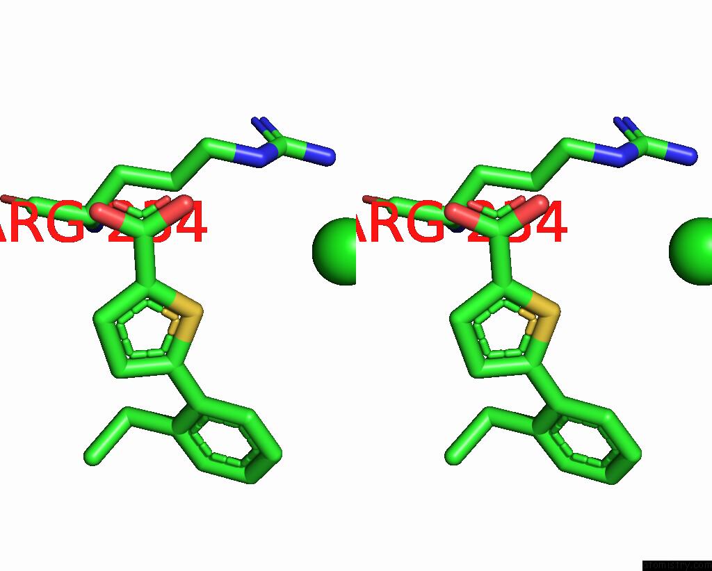
Chlorine binding site 1 out of 3 in 7mlt
Go back to
Chlorine binding site 1 out
of 3 in the Crystal Structure of Ricin A Chain in Complex with 5-(2-Ethylphenyl) Thiophene-2-Carboxylic Acid

Mono view

Stereo pair view
Mono view
Stereo pair view
A full contact list of Chlorine with other atoms in the Cl binding
site number 1 of Crystal Structure of Ricin A Chain in Complex with 5-(2-Ethylphenyl) Thiophene-2-Carboxylic Acid within 5.0Å range:
|
Chlorine binding site 2 out of 3 in 7mlt
Go back to
Chlorine binding site 2 out
of 3 in the Crystal Structure of Ricin A Chain in Complex with 5-(2-Ethylphenyl) Thiophene-2-Carboxylic Acid

Mono view
Stereo pair view
Mono view
Stereo pair view
A full contact list of Chlorine with other atoms in the Cl binding
site number 2 of Crystal Structure of Ricin A Chain in Complex with 5-(2-Ethylphenyl) Thiophene-2-Carboxylic Acid within 5.0Å range:
|
Chlorine binding site 3 out of 3 in 7mlt
Go back to
Chlorine binding site 3 out
of 3 in the Crystal Structure of Ricin A Chain in Complex with 5-(2-Ethylphenyl) Thiophene-2-Carboxylic Acid

Mono view

Stereo pair view
Mono view
Stereo pair view
A full contact list of Chlorine with other atoms in the Cl binding
site number 3 of Crystal Structure of Ricin A Chain in Complex with 5-(2-Ethylphenyl) Thiophene-2-Carboxylic Acid within 5.0Å range:
|
Reference:
X.P.Li,
R.K.Harijan,
B.Cao,
J.N.Kahn,
M.Pierce,
A.M.Tsymbal,
J.Y.Roberge,
D.Augeri,
N.E.Tumer.
Synthesis and Structural Characterization of Ricin Inhibitors Targeting Ribosome Binding Using Fragment-Based Methods and Structure-Based Design. J.Med.Chem. V. 64 15334 2021.
ISSN: ISSN 0022-2623
PubMed: 34648707
DOI: 10.1021/ACS.JMEDCHEM.1C01370
Page generated: Tue Jul 30 00:33:04 2024
ISSN: ISSN 0022-2623
PubMed: 34648707
DOI: 10.1021/ACS.JMEDCHEM.1C01370
Last articles
Zn in 9MJ5Zn in 9HNW
Zn in 9G0L
Zn in 9FNE
Zn in 9DZN
Zn in 9E0I
Zn in 9D32
Zn in 9DAK
Zn in 8ZXC
Zn in 8ZUF