Chlorine in PDB 7mov: PTP1B 1-301 F225Y-R199N Mutations
Enzymatic activity of PTP1B 1-301 F225Y-R199N Mutations
All present enzymatic activity of PTP1B 1-301 F225Y-R199N Mutations:
3.1.3.48;
3.1.3.48;
Protein crystallography data
The structure of PTP1B 1-301 F225Y-R199N Mutations, PDB code: 7mov
was solved by
K.R.Torgeson,
R.Page,
W.Peti,
with X-Ray Crystallography technique. A brief refinement statistics is given in the table below:
| Resolution Low / High (Å) | 37.82 / 1.65 |
| Space group | C 1 2 1 |
| Cell size a, b, c (Å), α, β, γ (°) | 114.359, 90.102, 74.49, 90, 110.93, 90 |
| R / Rfree (%) | 16.5 / 19.3 |
Chlorine Binding Sites:
The binding sites of Chlorine atom in the PTP1B 1-301 F225Y-R199N Mutations
(pdb code 7mov). This binding sites where shown within
5.0 Angstroms radius around Chlorine atom.
In total 3 binding sites of Chlorine where determined in the PTP1B 1-301 F225Y-R199N Mutations, PDB code: 7mov:
Jump to Chlorine binding site number: 1; 2; 3;
In total 3 binding sites of Chlorine where determined in the PTP1B 1-301 F225Y-R199N Mutations, PDB code: 7mov:
Jump to Chlorine binding site number: 1; 2; 3;
Chlorine binding site 1 out of 3 in 7mov
Go back to
Chlorine binding site 1 out
of 3 in the PTP1B 1-301 F225Y-R199N Mutations
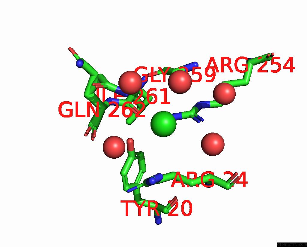
Mono view

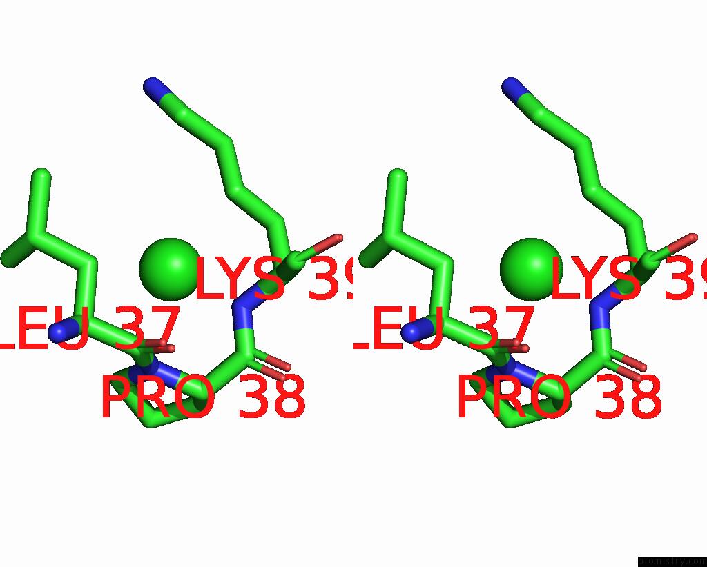
Stereo pair view
Mono view
Stereo pair view
A full contact list of Chlorine with other atoms in the Cl binding
site number 1 of PTP1B 1-301 F225Y-R199N Mutations within 5.0Å range:
|
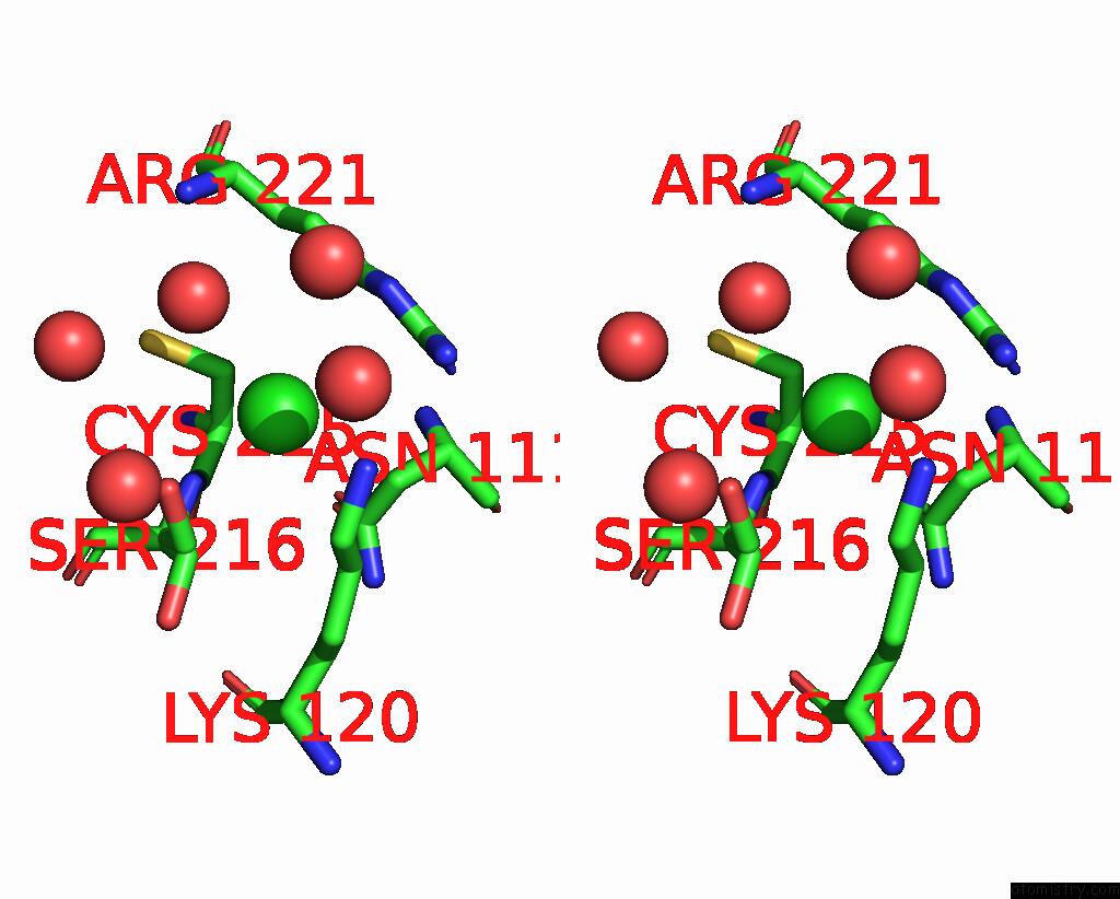
Chlorine binding site 2 out of 3 in 7mov
Go back to
Chlorine binding site 2 out
of 3 in the PTP1B 1-301 F225Y-R199N Mutations
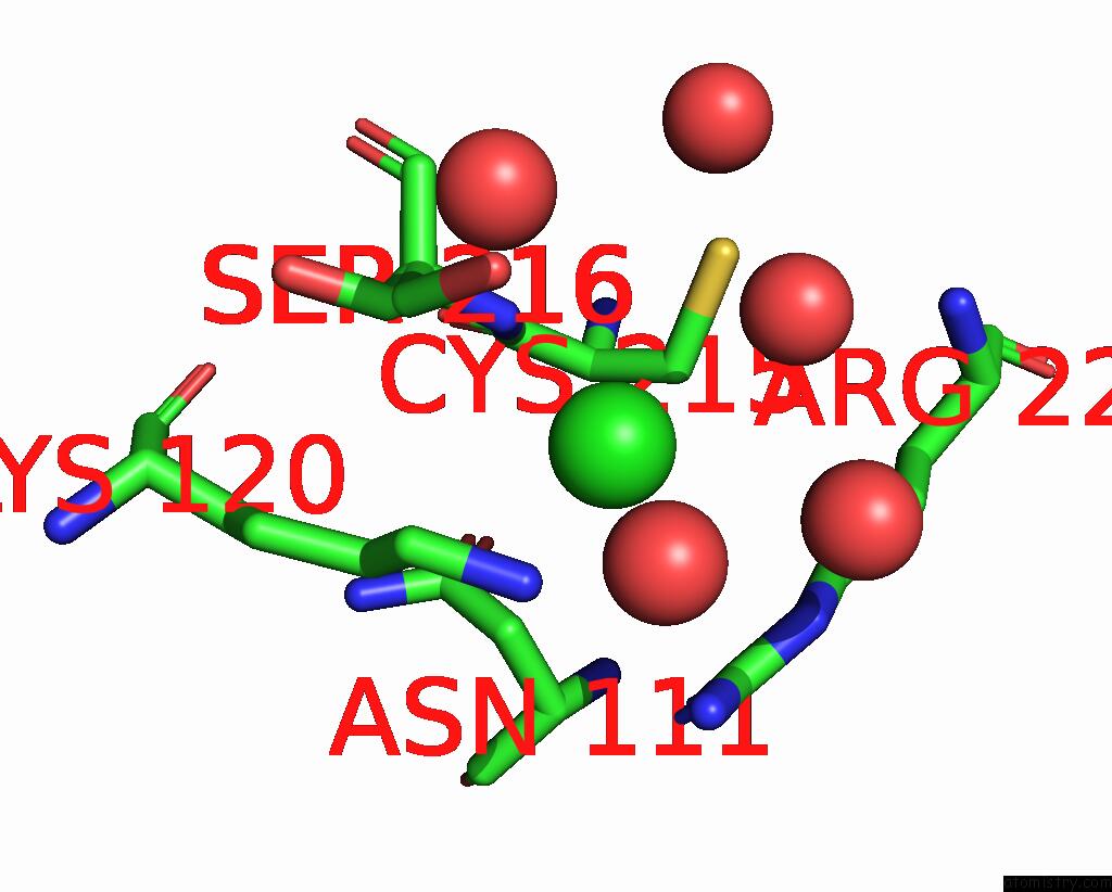
Mono view
Stereo pair view
Mono view
Stereo pair view
A full contact list of Chlorine with other atoms in the Cl binding
site number 2 of PTP1B 1-301 F225Y-R199N Mutations within 5.0Å range:
|
Chlorine binding site 3 out of 3 in 7mov
Go back to
Chlorine binding site 3 out
of 3 in the PTP1B 1-301 F225Y-R199N Mutations
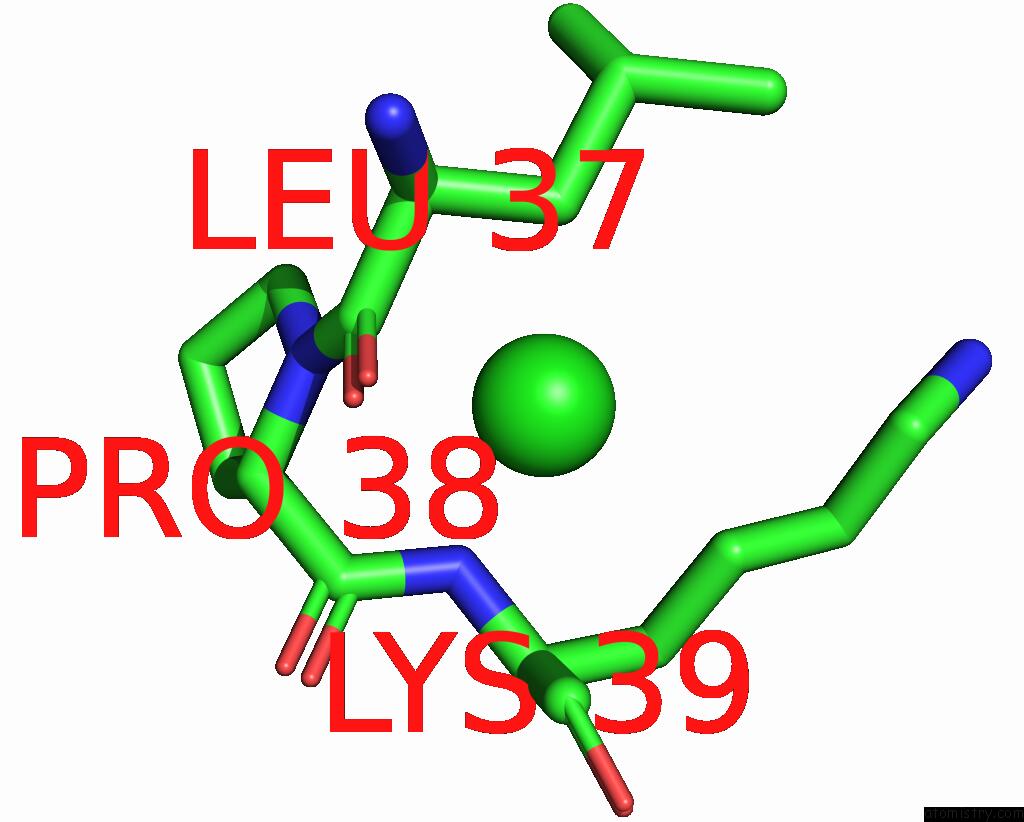
Mono view
Stereo pair view
Mono view
Stereo pair view
A full contact list of Chlorine with other atoms in the Cl binding
site number 3 of PTP1B 1-301 F225Y-R199N Mutations within 5.0Å range:
|
Reference:
K.R.Torgeson,
M.W.Clarkson,
D.Granata,
K.Lindorff-Larsen,
R.Page,
W.Peti.
Conserved Conformational Dynamics Determine Enzyme Activity. Sci Adv V. 8 O5546 2022.
ISSN: ESSN 2375-2548
PubMed: 35921420
DOI: 10.1126/SCIADV.ABO5546
Page generated: Tue Jul 30 00:36:13 2024
ISSN: ESSN 2375-2548
PubMed: 35921420
DOI: 10.1126/SCIADV.ABO5546
Last articles
Zn in 9MJ5Zn in 9HNW
Zn in 9G0L
Zn in 9FNE
Zn in 9DZN
Zn in 9E0I
Zn in 9D32
Zn in 9DAK
Zn in 8ZXC
Zn in 8ZUF