Chlorine in PDB 7p2b: Crystal Structure of Human Gelsolin Amyloid Mutant A551P
Protein crystallography data
The structure of Crystal Structure of Human Gelsolin Amyloid Mutant A551P, PDB code: 7p2b
was solved by
M.Bollati,
M.De Rosa,
with X-Ray Crystallography technique. A brief refinement statistics is given in the table below:
| Resolution Low / High (Å) | 19.91 / 3.00 |
| Space group | P 4 21 2 |
| Cell size a, b, c (Å), α, β, γ (°) | 170.07, 170.07, 152, 90, 90, 90 |
| R / Rfree (%) | 21.3 / 26.3 |
Chlorine Binding Sites:
The binding sites of Chlorine atom in the Crystal Structure of Human Gelsolin Amyloid Mutant A551P
(pdb code 7p2b). This binding sites where shown within
5.0 Angstroms radius around Chlorine atom.
In total only one binding site of Chlorine was determined in the Crystal Structure of Human Gelsolin Amyloid Mutant A551P, PDB code: 7p2b:
In total only one binding site of Chlorine was determined in the Crystal Structure of Human Gelsolin Amyloid Mutant A551P, PDB code: 7p2b:
Chlorine binding site 1 out of 1 in 7p2b
Go back to
Chlorine binding site 1 out
of 1 in the Crystal Structure of Human Gelsolin Amyloid Mutant A551P
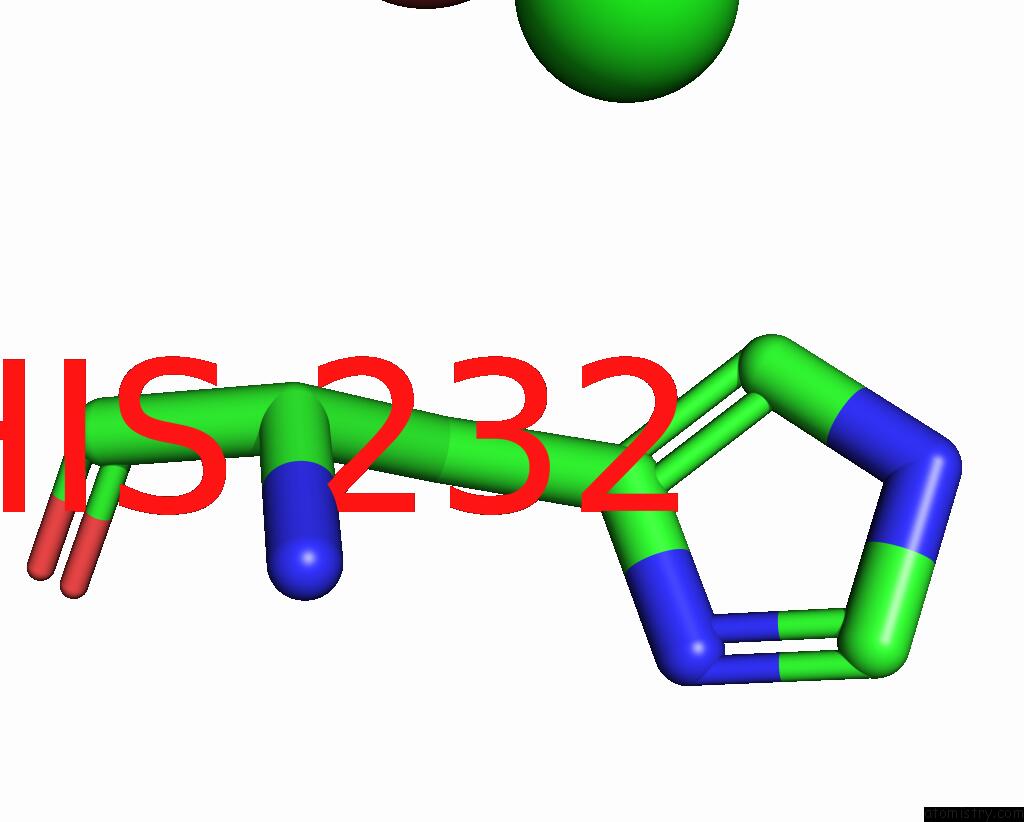
Mono view
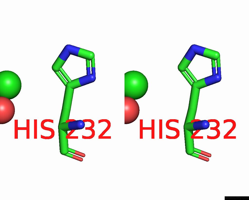
Stereo pair view
Mono view
Stereo pair view
A full contact list of Chlorine with other atoms in the Cl binding
site number 1 of Crystal Structure of Human Gelsolin Amyloid Mutant A551P within 5.0Å range:
|
Reference:
M.Bollati,
L.Diomede,
T.Giorgino,
C.Natale,
E.Fagnani,
I.Boniardi,
A.Barbiroli,
R.Alemani,
M.Beeg,
M.Gobbi,
A.Fakin,
E.Mastrangelo,
M.Milani,
G.Presciuttini,
E.Gabellieri,
P.Cioni,
M.De Rosa.
A Novel Hotspot of Gelsolin Instability Triggers An Alternative Mechanism of Amyloid Aggregation. Comput Struct Biotechnol J V. 19 6355 2021.
ISSN: ESSN 2001-0370
PubMed: 34938411
DOI: 10.1016/J.CSBJ.2021.11.025
Page generated: Tue Jul 30 02:14:37 2024
ISSN: ESSN 2001-0370
PubMed: 34938411
DOI: 10.1016/J.CSBJ.2021.11.025
Last articles
Zn in 9JYWZn in 9IR4
Zn in 9IR3
Zn in 9GMX
Zn in 9GMW
Zn in 9JEJ
Zn in 9ERF
Zn in 9ERE
Zn in 9EGV
Zn in 9EGW