Chlorine in PDB 7pyf: Structure of Lpmo in Complex with Cellotetraose at 1.39X10^5 Gy
Protein crystallography data
The structure of Structure of Lpmo in Complex with Cellotetraose at 1.39X10^5 Gy, PDB code: 7pyf
was solved by
T.Tandrup,
L.Lo Leggio,
with X-Ray Crystallography technique. A brief refinement statistics is given in the table below:
| Resolution Low / High (Å) | 44.45 / 1.90 |
| Space group | P 41 3 2 |
| Cell size a, b, c (Å), α, β, γ (°) | 125.62, 125.62, 125.62, 90, 90, 90 |
| R / Rfree (%) | 25.4 / 28.7 |
Other elements in 7pyf:
The structure of Structure of Lpmo in Complex with Cellotetraose at 1.39X10^5 Gy also contains other interesting chemical elements:
| Copper | (Cu) | 1 atom |
Chlorine Binding Sites:
The binding sites of Chlorine atom in the Structure of Lpmo in Complex with Cellotetraose at 1.39X10^5 Gy
(pdb code 7pyf). This binding sites where shown within
5.0 Angstroms radius around Chlorine atom.
In total 3 binding sites of Chlorine where determined in the Structure of Lpmo in Complex with Cellotetraose at 1.39X10^5 Gy, PDB code: 7pyf:
Jump to Chlorine binding site number: 1; 2; 3;
In total 3 binding sites of Chlorine where determined in the Structure of Lpmo in Complex with Cellotetraose at 1.39X10^5 Gy, PDB code: 7pyf:
Jump to Chlorine binding site number: 1; 2; 3;
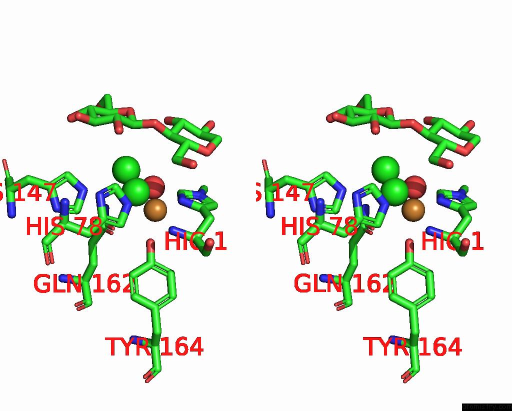
Chlorine binding site 1 out of 3 in 7pyf
Go back to
Chlorine binding site 1 out
of 3 in the Structure of Lpmo in Complex with Cellotetraose at 1.39X10^5 Gy
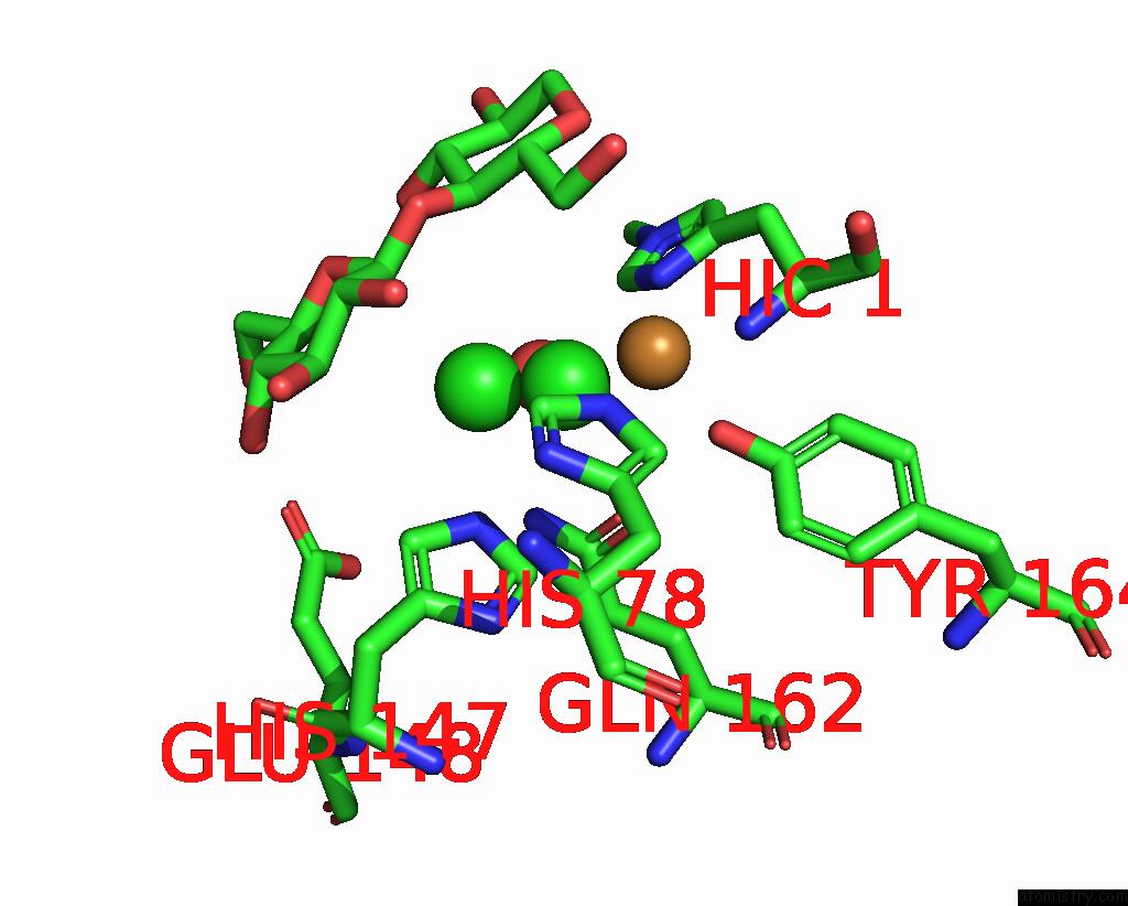
Mono view

Stereo pair view
Mono view
Stereo pair view
A full contact list of Chlorine with other atoms in the Cl binding
site number 1 of Structure of Lpmo in Complex with Cellotetraose at 1.39X10^5 Gy within 5.0Å range:
|
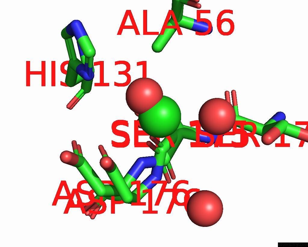
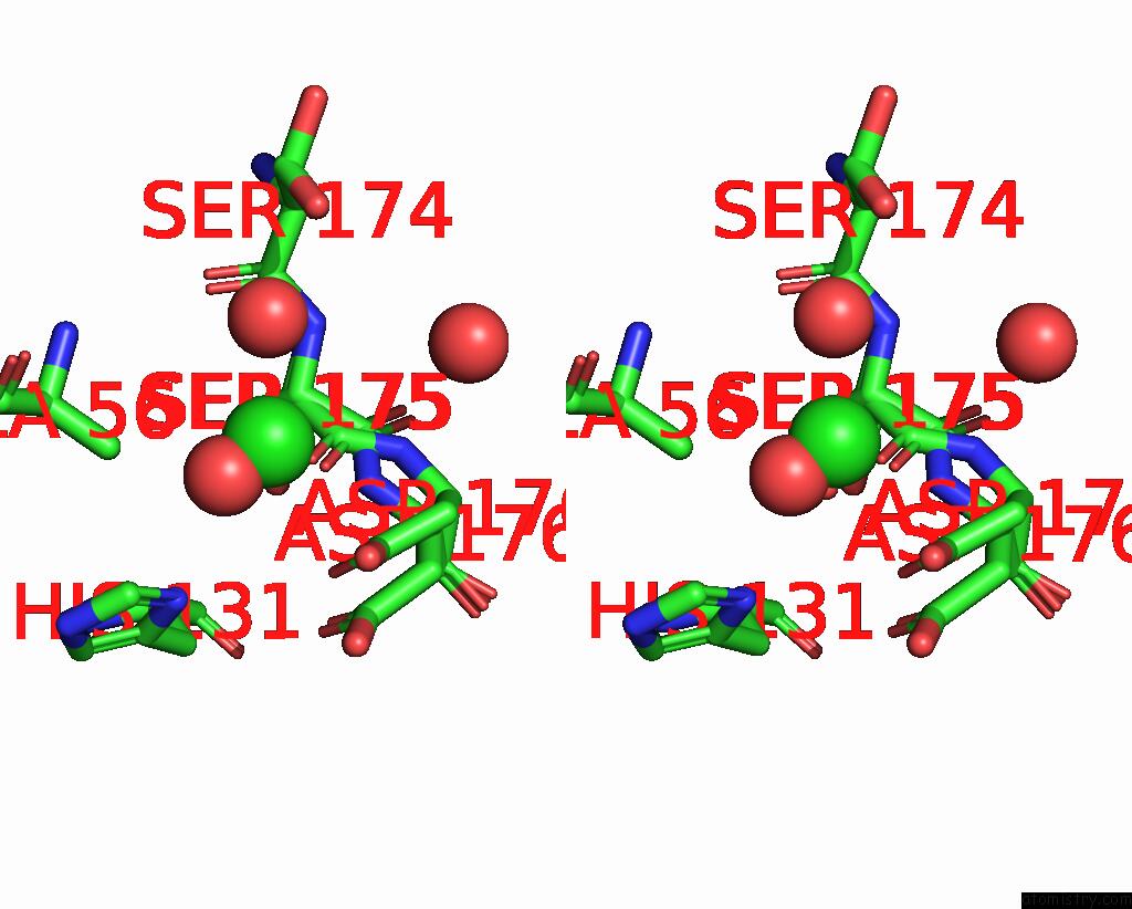
Chlorine binding site 2 out of 3 in 7pyf
Go back to
Chlorine binding site 2 out
of 3 in the Structure of Lpmo in Complex with Cellotetraose at 1.39X10^5 Gy

Mono view
Stereo pair view
Mono view
Stereo pair view
A full contact list of Chlorine with other atoms in the Cl binding
site number 2 of Structure of Lpmo in Complex with Cellotetraose at 1.39X10^5 Gy within 5.0Å range:
|
Chlorine binding site 3 out of 3 in 7pyf
Go back to
Chlorine binding site 3 out
of 3 in the Structure of Lpmo in Complex with Cellotetraose at 1.39X10^5 Gy
Mono view
Stereo pair view
Mono view
Stereo pair view
A full contact list of Chlorine with other atoms in the Cl binding
site number 3 of Structure of Lpmo in Complex with Cellotetraose at 1.39X10^5 Gy within 5.0Å range:
|
Reference:
T.Tandrup,
S.J.Muderspach,
S.Banerjee,
G.Santoni,
J.O.Ipsen,
C.Hernandez-Rollan,
M.H.H.Norholm,
K.S.Johansen,
F.Meilleur,
L.Lo Leggio.
Changes in Active-Site Geometry on X-Ray Photoreduction of A Lytic Polysaccharide Monooxygenase Active-Site Copper and Saccharide Binding. Iucrj V. 9 666 2022.
ISSN: ESSN 2052-2525
PubMed: 36071795
DOI: 10.1107/S2052252522007175
Page generated: Tue Jul 30 02:54:15 2024
ISSN: ESSN 2052-2525
PubMed: 36071795
DOI: 10.1107/S2052252522007175
Last articles
Zn in 9J0NZn in 9J0O
Zn in 9J0P
Zn in 9FJX
Zn in 9EKB
Zn in 9C0F
Zn in 9CAH
Zn in 9CH0
Zn in 9CH3
Zn in 9CH1