Chlorine in PDB 7te2: Crystal Structure of Aerr From Rhodobacter Capsulatus at 2.25 A.
Protein crystallography data
The structure of Crystal Structure of Aerr From Rhodobacter Capsulatus at 2.25 A., PDB code: 7te2
was solved by
V.Dragnea,
G.Gonzalez-Gutierrez,
C.E.Bauer,
with X-Ray Crystallography technique. A brief refinement statistics is given in the table below:
| Resolution Low / High (Å) | 47.32 / 2.25 |
| Space group | P 61 2 2 |
| Cell size a, b, c (Å), α, β, γ (°) | 63.939, 63.939, 182.298, 90, 90, 120 |
| R / Rfree (%) | 23 / 25.9 |
Other elements in 7te2:
The structure of Crystal Structure of Aerr From Rhodobacter Capsulatus at 2.25 A. also contains other interesting chemical elements:
| Cobalt | (Co) | 1 atom |
Chlorine Binding Sites:
The binding sites of Chlorine atom in the Crystal Structure of Aerr From Rhodobacter Capsulatus at 2.25 A.
(pdb code 7te2). This binding sites where shown within
5.0 Angstroms radius around Chlorine atom.
In total 3 binding sites of Chlorine where determined in the Crystal Structure of Aerr From Rhodobacter Capsulatus at 2.25 A., PDB code: 7te2:
Jump to Chlorine binding site number: 1; 2; 3;
In total 3 binding sites of Chlorine where determined in the Crystal Structure of Aerr From Rhodobacter Capsulatus at 2.25 A., PDB code: 7te2:
Jump to Chlorine binding site number: 1; 2; 3;
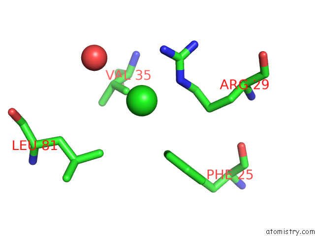
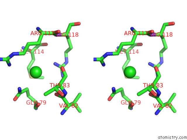
Chlorine binding site 1 out of 3 in 7te2
Go back to
Chlorine binding site 1 out
of 3 in the Crystal Structure of Aerr From Rhodobacter Capsulatus at 2.25 A.
Mono view
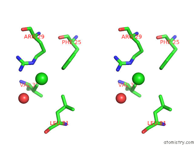
Stereo pair view
Mono view
Stereo pair view
A full contact list of Chlorine with other atoms in the Cl binding
site number 1 of Crystal Structure of Aerr From Rhodobacter Capsulatus at 2.25 A. within 5.0Å range:
|
Chlorine binding site 2 out of 3 in 7te2
Go back to
Chlorine binding site 2 out
of 3 in the Crystal Structure of Aerr From Rhodobacter Capsulatus at 2.25 A.

Mono view
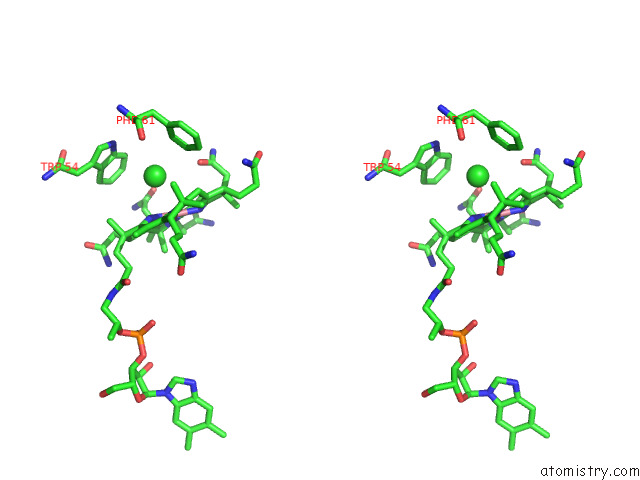
Stereo pair view
Mono view
Stereo pair view
A full contact list of Chlorine with other atoms in the Cl binding
site number 2 of Crystal Structure of Aerr From Rhodobacter Capsulatus at 2.25 A. within 5.0Å range:
|
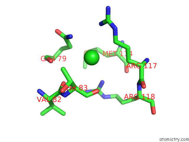
Chlorine binding site 3 out of 3 in 7te2
Go back to
Chlorine binding site 3 out
of 3 in the Crystal Structure of Aerr From Rhodobacter Capsulatus at 2.25 A.
Mono view
Stereo pair view
Mono view
Stereo pair view
A full contact list of Chlorine with other atoms in the Cl binding
site number 3 of Crystal Structure of Aerr From Rhodobacter Capsulatus at 2.25 A. within 5.0Å range:
|
Reference:
V.Dragnea,
G.Gonzalez-Gutierrez,
C.E.Bauer.
Structural Analyses of Crtj and Its B 12 -Binding Co-Regulators Saerr and Laerr From the Purple Photosynthetic Bacterium Rhodobacter Capsulatus. Microorganisms V. 10 2022.
ISSN: ESSN 2076-2607
PubMed: 35630357
DOI: 10.3390/MICROORGANISMS10050912
Page generated: Tue Jul 30 04:33:49 2024
ISSN: ESSN 2076-2607
PubMed: 35630357
DOI: 10.3390/MICROORGANISMS10050912
Last articles
Zn in 9JYWZn in 9IR4
Zn in 9IR3
Zn in 9GMX
Zn in 9GMW
Zn in 9JEJ
Zn in 9ERF
Zn in 9ERE
Zn in 9EGV
Zn in 9EGW