Chlorine in PDB 7toj: Crystal Structure of Carbohydrate Esterase Cspacxe, Apoenzyme
Protein crystallography data
The structure of Crystal Structure of Carbohydrate Esterase Cspacxe, Apoenzyme, PDB code: 7toj
was solved by
P.J.Stogios,
T.Skarina,
R.Di Leo,
E.Jurak,
E.Master,
with X-Ray Crystallography technique. A brief refinement statistics is given in the table below:
Resolution Low / High (Å)
|
35.36 /
1.30
|
Space group
|
C 1 2 1
|
Cell size a, b, c (Å), α, β, γ (°)
|
120.16,
77.31,
42.08,
90,
96.89,
90
|
R / Rfree (%)
|
14.1 /
16.4
|
Chlorine Binding Sites:
The binding sites of Chlorine atom in the Crystal Structure of Carbohydrate Esterase Cspacxe, Apoenzyme
(pdb code 7toj). This binding sites where shown within
5.0 Angstroms radius around Chlorine atom.
In total 3 binding sites of Chlorine where determined in the
Crystal Structure of Carbohydrate Esterase Cspacxe, Apoenzyme, PDB code: 7toj:
Jump to Chlorine binding site number:
1;
2;
3;
Chlorine binding site 1 out
of 3 in 7toj
Go back to
Chlorine Binding Sites List in 7toj
Chlorine binding site 1 out
of 3 in the Crystal Structure of Carbohydrate Esterase Cspacxe, Apoenzyme
Mono view
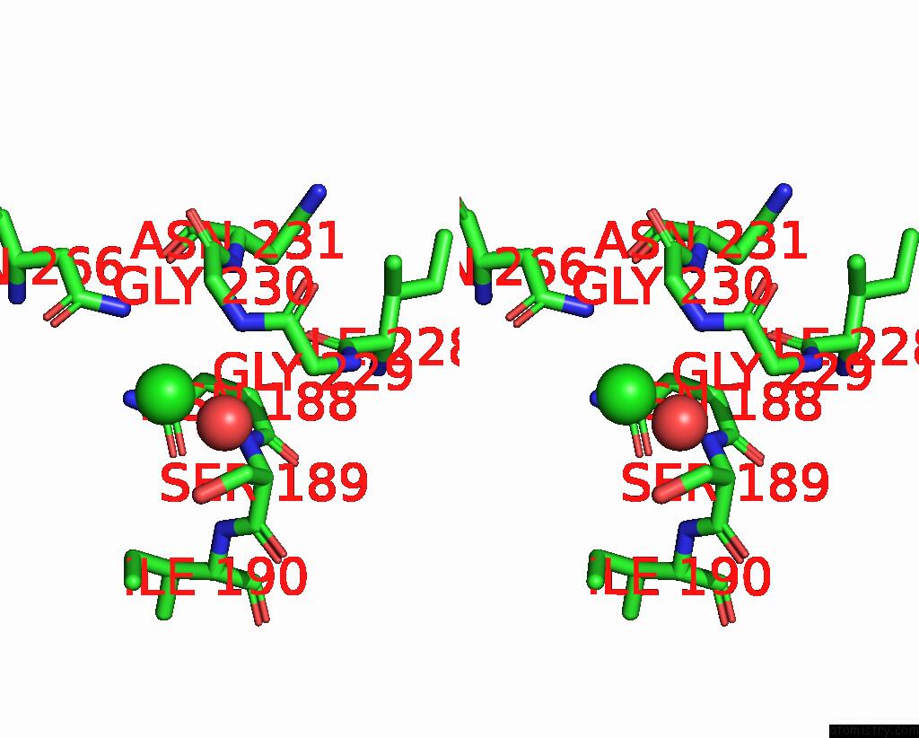
Stereo pair view
|
A full contact list of Chlorine with other atoms in the Cl binding
site number 1 of Crystal Structure of Carbohydrate Esterase Cspacxe, Apoenzyme within 5.0Å range:
probe
|
atom
|
residue
|
distance (Å)
|
B
|
Occ
|
A:Cl401
b:22.7
occ:0.92
|
N
|
A:GLY230
|
3.1
|
22.9
|
1.0
|
OG
|
A:SER189
|
3.2
|
24.6
|
1.0
|
ND2
|
A:ASN266
|
3.2
|
21.1
|
1.0
|
N
|
A:SER189
|
3.2
|
18.4
|
1.0
|
CB
|
A:SER189
|
3.3
|
22.1
|
1.0
|
CB
|
A:ASN188
|
3.6
|
18.4
|
1.0
|
CA
|
A:GLY230
|
3.7
|
24.4
|
1.0
|
CG
|
A:ASN188
|
3.8
|
20.7
|
1.0
|
CA
|
A:SER189
|
3.9
|
18.2
|
1.0
|
OD1
|
A:ASN266
|
4.0
|
26.5
|
1.0
|
CG
|
A:ASN266
|
4.1
|
21.4
|
1.0
|
O
|
A:HOH920
|
4.2
|
47.7
|
1.0
|
C
|
A:GLY229
|
4.2
|
23.1
|
1.0
|
ND2
|
A:ASN188
|
4.2
|
22.3
|
1.0
|
OD1
|
A:ASN188
|
4.2
|
20.6
|
1.0
|
C
|
A:ASN188
|
4.3
|
16.9
|
1.0
|
CA
|
A:GLY229
|
4.4
|
21.8
|
1.0
|
C
|
A:GLY230
|
4.5
|
22.2
|
1.0
|
CA
|
A:ASN188
|
4.5
|
16.3
|
1.0
|
N
|
A:ASN231
|
4.6
|
19.7
|
1.0
|
C
|
A:SER189
|
4.8
|
17.5
|
1.0
|
N
|
A:ILE190
|
4.9
|
18.0
|
1.0
|
O
|
A:ILE228
|
5.0
|
17.6
|
1.0
|
|
Chlorine binding site 2 out
of 3 in 7toj
Go back to
Chlorine Binding Sites List in 7toj
Chlorine binding site 2 out
of 3 in the Crystal Structure of Carbohydrate Esterase Cspacxe, Apoenzyme
Mono view
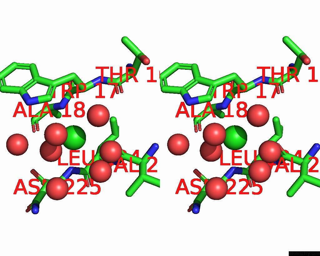
Stereo pair view
|
A full contact list of Chlorine with other atoms in the Cl binding
site number 2 of Crystal Structure of Carbohydrate Esterase Cspacxe, Apoenzyme within 5.0Å range:
probe
|
atom
|
residue
|
distance (Å)
|
B
|
Occ
|
A:Cl402
b:18.9
occ:0.52
|
O
|
A:HOH924
|
2.7
|
18.6
|
1.0
|
N
|
A:ASN225
|
3.0
|
14.6
|
1.0
|
O
|
A:VAL223
|
3.0
|
16.9
|
1.0
|
N
|
A:ALA18
|
3.1
|
14.2
|
1.0
|
O
|
A:ALA18
|
3.2
|
15.0
|
1.0
|
CA
|
A:LEU224
|
3.3
|
14.2
|
1.0
|
O
|
A:HOH711
|
3.5
|
20.0
|
1.0
|
C
|
A:LEU224
|
3.6
|
13.9
|
1.0
|
CA
|
A:TRP17
|
3.6
|
13.9
|
1.0
|
CD1
|
A:TRP17
|
3.8
|
15.7
|
1.0
|
C
|
A:TRP17
|
3.8
|
13.9
|
1.0
|
C
|
A:VAL223
|
3.9
|
15.3
|
1.0
|
CB
|
A:ASN225
|
3.9
|
15.9
|
1.0
|
C
|
A:ALA18
|
4.0
|
14.2
|
1.0
|
CA
|
A:ASN225
|
4.0
|
15.1
|
1.0
|
N
|
A:LEU224
|
4.0
|
15.2
|
1.0
|
CA
|
A:ALA18
|
4.1
|
14.2
|
1.0
|
O
|
A:HOH613
|
4.1
|
17.8
|
1.0
|
CG
|
A:TRP17
|
4.2
|
14.3
|
1.0
|
O
|
A:HOH679
|
4.2
|
22.4
|
0.6
|
O
|
A:HOH679
|
4.3
|
30.1
|
0.4
|
NE1
|
A:TRP17
|
4.3
|
15.8
|
1.0
|
CD2
|
A:LEU224
|
4.4
|
17.3
|
1.0
|
CB
|
A:TRP17
|
4.4
|
15.0
|
1.0
|
CB
|
A:LEU224
|
4.4
|
15.9
|
1.0
|
CB
|
A:ALA18
|
4.6
|
15.9
|
1.0
|
O
|
A:HOH563
|
4.7
|
19.0
|
1.0
|
O
|
A:THR16
|
4.7
|
14.9
|
1.0
|
N
|
A:TRP17
|
4.7
|
14.1
|
1.0
|
O
|
A:HOH785
|
4.8
|
19.6
|
1.0
|
O
|
A:ASN225
|
4.8
|
15.6
|
1.0
|
O
|
A:LEU224
|
4.8
|
15.0
|
1.0
|
C
|
A:ASN225
|
4.9
|
15.1
|
1.0
|
CD2
|
A:TRP17
|
4.9
|
14.3
|
1.0
|
|
Chlorine binding site 3 out
of 3 in 7toj
Go back to
Chlorine Binding Sites List in 7toj
Chlorine binding site 3 out
of 3 in the Crystal Structure of Carbohydrate Esterase Cspacxe, Apoenzyme
Mono view
Stereo pair view
|
A full contact list of Chlorine with other atoms in the Cl binding
site number 3 of Crystal Structure of Carbohydrate Esterase Cspacxe, Apoenzyme within 5.0Å range:
probe
|
atom
|
residue
|
distance (Å)
|
B
|
Occ
|
A:Cl403
b:20.5
occ:0.54
|
OG
|
A:SER144
|
2.8
|
18.0
|
1.0
|
O
|
A:THR143
|
2.9
|
19.4
|
1.0
|
N
|
A:ILE43
|
2.9
|
16.3
|
1.0
|
NE1
|
A:TRP166
|
3.1
|
16.4
|
1.0
|
C
|
A:THR143
|
3.4
|
18.4
|
1.0
|
CA
|
A:GLN42
|
3.4
|
16.8
|
1.0
|
CG2
|
A:THR142
|
3.4
|
17.9
|
1.0
|
CD1
|
A:TRP166
|
3.5
|
16.7
|
1.0
|
C
|
A:GLN42
|
3.6
|
16.2
|
1.0
|
N
|
A:SER144
|
3.6
|
19.1
|
1.0
|
O
|
A:ILE43
|
3.6
|
16.7
|
1.0
|
CA
|
A:SER144
|
3.7
|
19.6
|
1.0
|
CB
|
A:SER144
|
3.8
|
17.6
|
1.0
|
CB
|
A:GLN42
|
3.8
|
17.0
|
1.0
|
O
|
A:THR142
|
3.9
|
19.7
|
1.0
|
CA
|
A:ILE43
|
3.9
|
16.9
|
1.0
|
CB
|
A:THR142
|
3.9
|
17.5
|
1.0
|
C
|
A:THR142
|
4.1
|
18.9
|
1.0
|
CB
|
A:ILE43
|
4.1
|
18.0
|
1.0
|
C
|
A:ILE43
|
4.2
|
16.1
|
1.0
|
CG1
|
A:ILE43
|
4.3
|
22.3
|
1.0
|
CE2
|
A:TRP166
|
4.3
|
15.0
|
1.0
|
N
|
A:THR143
|
4.4
|
18.8
|
1.0
|
CG
|
A:GLN42
|
4.4
|
17.5
|
1.0
|
CA
|
A:THR143
|
4.5
|
19.1
|
1.0
|
O
|
A:ARG41
|
4.5
|
19.1
|
1.0
|
N
|
A:GLN42
|
4.6
|
16.4
|
1.0
|
CA
|
A:THR142
|
4.7
|
17.7
|
1.0
|
O
|
A:HOH636
|
4.7
|
21.8
|
1.0
|
O
|
A:GLN42
|
4.8
|
18.0
|
1.0
|
CG
|
A:TRP166
|
4.8
|
16.4
|
1.0
|
O
|
A:HIS165
|
4.9
|
19.1
|
1.0
|
|
Reference:
L.Penttinen,
V.Kouhi,
R.Faure,
T.Skarina,
P.Stogios,
E.Master,
E.Jurak.
Elucidating Sequence and Structural Determinants of Carbohydrate Esterases For Complete Deacetylation of Substituted Xylans. Molecules V. 27 2022.
ISSN: ESSN 1420-3049
PubMed: 35566004
DOI: 10.3390/MOLECULES27092655
Page generated: Tue Jul 30 04:45:42 2024
|