Chlorine in PDB 7vgv: Anion Free Form of Light-Driven Chloride Ion-Pumping Rhodopsin, Nm-R3, Structure Determined By Serial Femtosecond Crystallography at Sacla
Protein crystallography data
The structure of Anion Free Form of Light-Driven Chloride Ion-Pumping Rhodopsin, Nm-R3, Structure Determined By Serial Femtosecond Crystallography at Sacla, PDB code: 7vgv
was solved by
T.Hosaka,
E.Nango,
T.Nakane,
F.Luo,
T.Kimura-Someya,
M.Shirouzu,
with X-Ray Crystallography technique. A brief refinement statistics is given in the table below:
| Resolution Low / High (Å) | 44.46 / 2.30 |
| Space group | P 21 21 21 |
| Cell size a, b, c (Å), α, β, γ (°) | 68.4, 69.5, 231.4, 90, 90, 90 |
| R / Rfree (%) | 21 / 24.1 |
Chlorine Binding Sites:
The binding sites of Chlorine atom in the Anion Free Form of Light-Driven Chloride Ion-Pumping Rhodopsin, Nm-R3, Structure Determined By Serial Femtosecond Crystallography at Sacla
(pdb code 7vgv). This binding sites where shown within
5.0 Angstroms radius around Chlorine atom.
In total 2 binding sites of Chlorine where determined in the Anion Free Form of Light-Driven Chloride Ion-Pumping Rhodopsin, Nm-R3, Structure Determined By Serial Femtosecond Crystallography at Sacla, PDB code: 7vgv:
Jump to Chlorine binding site number: 1; 2;
In total 2 binding sites of Chlorine where determined in the Anion Free Form of Light-Driven Chloride Ion-Pumping Rhodopsin, Nm-R3, Structure Determined By Serial Femtosecond Crystallography at Sacla, PDB code: 7vgv:
Jump to Chlorine binding site number: 1; 2;
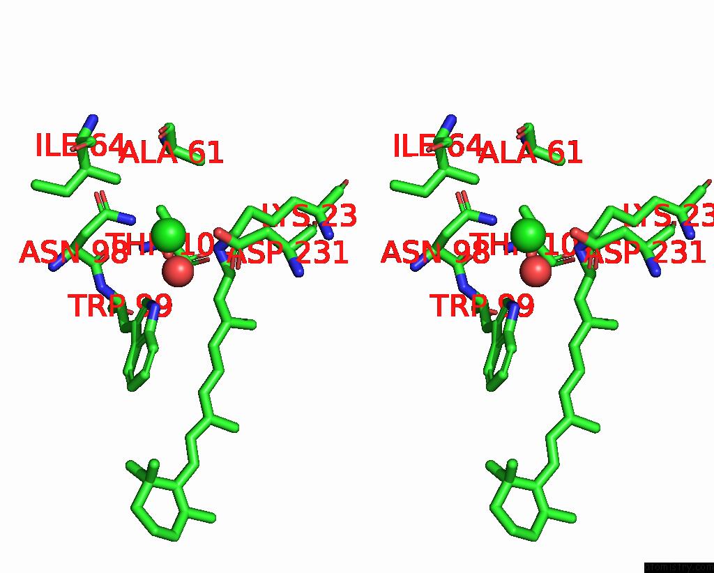
Chlorine binding site 1 out of 2 in 7vgv
Go back to
Chlorine binding site 1 out
of 2 in the Anion Free Form of Light-Driven Chloride Ion-Pumping Rhodopsin, Nm-R3, Structure Determined By Serial Femtosecond Crystallography at Sacla

Mono view
Stereo pair view
Mono view
Stereo pair view
A full contact list of Chlorine with other atoms in the Cl binding
site number 1 of Anion Free Form of Light-Driven Chloride Ion-Pumping Rhodopsin, Nm-R3, Structure Determined By Serial Femtosecond Crystallography at Sacla within 5.0Å range:
|
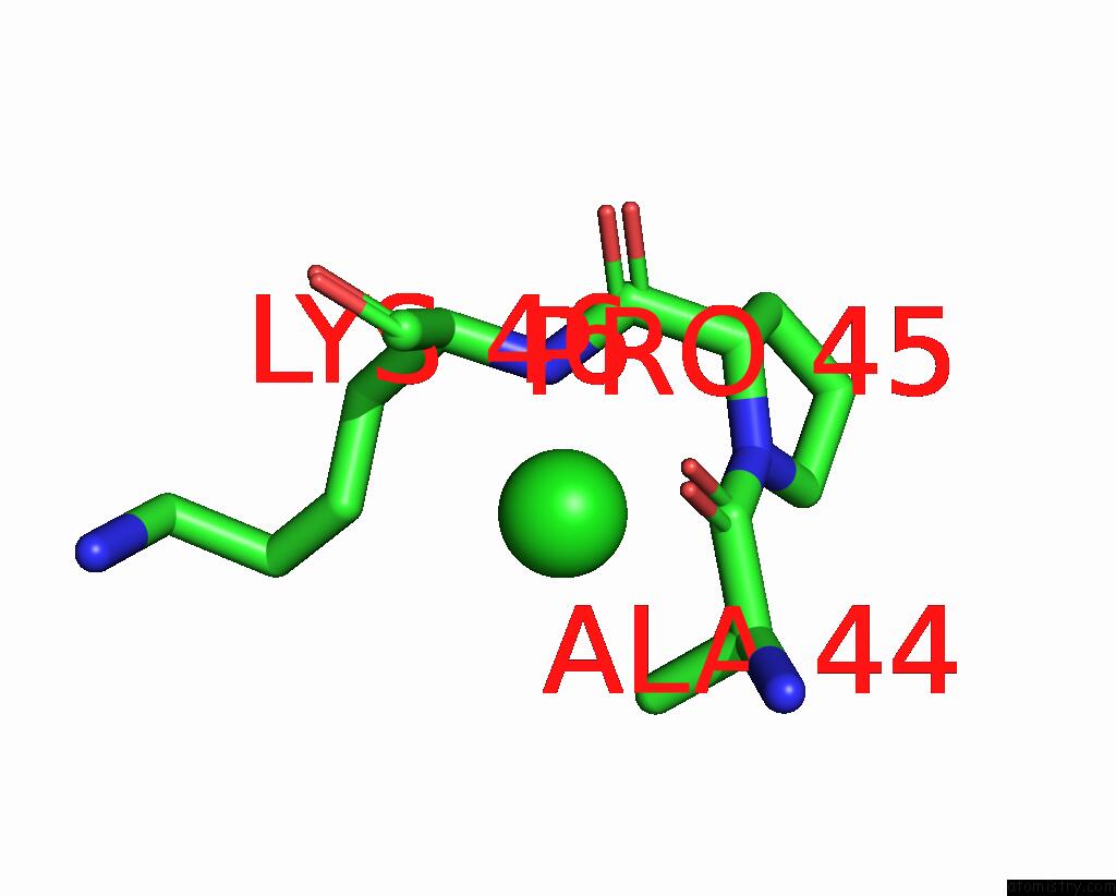
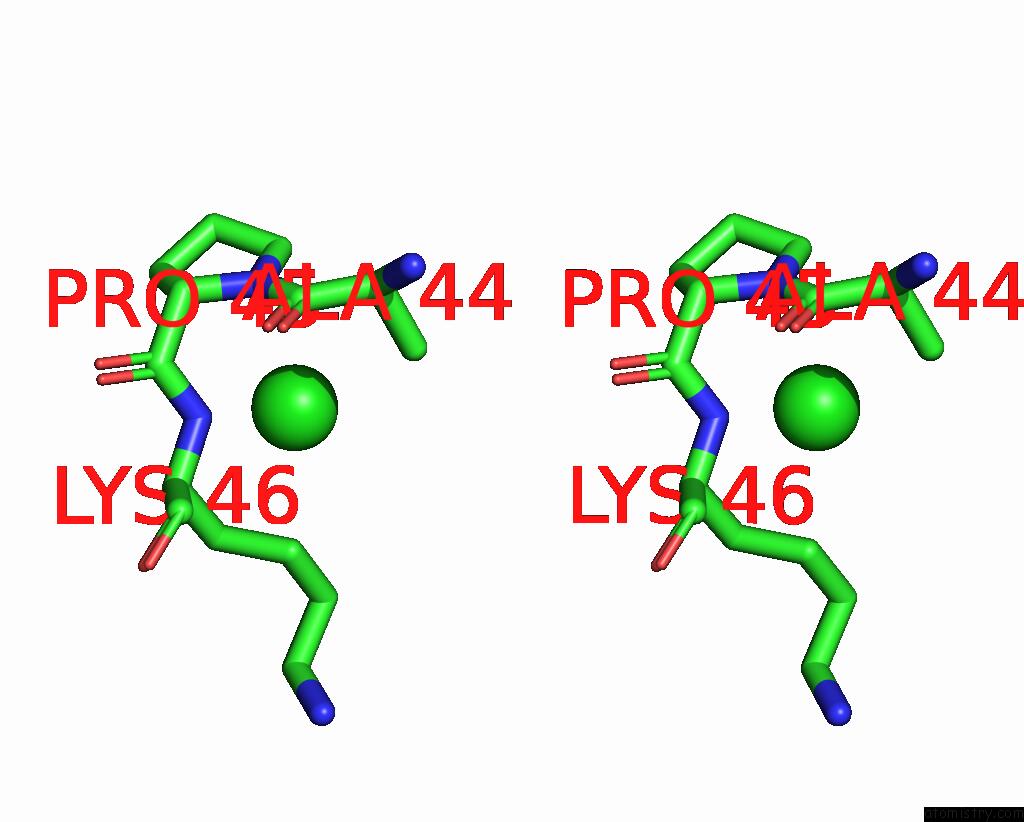
Chlorine binding site 2 out of 2 in 7vgv
Go back to
Chlorine binding site 2 out
of 2 in the Anion Free Form of Light-Driven Chloride Ion-Pumping Rhodopsin, Nm-R3, Structure Determined By Serial Femtosecond Crystallography at Sacla
Mono view
Stereo pair view
Mono view
Stereo pair view
A full contact list of Chlorine with other atoms in the Cl binding
site number 2 of Anion Free Form of Light-Driven Chloride Ion-Pumping Rhodopsin, Nm-R3, Structure Determined By Serial Femtosecond Crystallography at Sacla within 5.0Å range:
|
Reference:
T.Hosaka,
T.Nomura,
M.Kubo,
T.Nakane,
L.Fangjia,
S.I.Sekine,
T.Ito,
K.Murayama,
K.Ihara,
H.Ehara,
K.Kashiwagi,
K.Katsura,
R.Akasaka,
T.Hisano,
T.Tanaka,
R.Tanaka,
T.Arima,
A.Yamashita,
M.Sugahara,
H.Naitow,
Y.Matsuura,
S.Yoshizawa,
K.Tono,
S.Owada,
O.Nureki,
T.Kimura-Someya,
S.Iwata,
E.Nango,
M.Shirouzu.
Conformational Alterations in Unidirectional Ion Transport of A Light-Driven Chloride Pump Revealed Using X-Ray Free Electron Lasers. Proc.Natl.Acad.Sci.Usa V. 119 2022.
ISSN: ESSN 1091-6490
PubMed: 35197289
DOI: 10.1073/PNAS.2117433119
Page generated: Tue Jul 30 05:21:54 2024
ISSN: ESSN 1091-6490
PubMed: 35197289
DOI: 10.1073/PNAS.2117433119
Last articles
Zn in 9MJ5Zn in 9HNW
Zn in 9G0L
Zn in 9FNE
Zn in 9DZN
Zn in 9E0I
Zn in 9D32
Zn in 9DAK
Zn in 8ZXC
Zn in 8ZUF