Chlorine in PDB 7zsi: Structure of Orange Carotenoid Protein with Canthaxanthin Bound After 5 Minutes of Illumination
Protein crystallography data
The structure of Structure of Orange Carotenoid Protein with Canthaxanthin Bound After 5 Minutes of Illumination, PDB code: 7zsi
was solved by
V.U.Chukhutsina,
J.M.Baxter,
A.Fadini,
R.M.Morgan,
M.A.Pope,
K.Maghlaoui,
C.Orr,
A.Wagner,
J.J.Van Thor,
with X-Ray Crystallography technique. A brief refinement statistics is given in the table below:
| Resolution Low / High (Å) | 55.54 / 1.40 |
| Space group | P 32 2 1 |
| Cell size a, b, c (Å), α, β, γ (°) | 82.78, 82.78, 87.604, 90, 90, 120 |
| R / Rfree (%) | 19.1 / 20.5 |
Chlorine Binding Sites:
The binding sites of Chlorine atom in the Structure of Orange Carotenoid Protein with Canthaxanthin Bound After 5 Minutes of Illumination
(pdb code 7zsi). This binding sites where shown within
5.0 Angstroms radius around Chlorine atom.
In total 2 binding sites of Chlorine where determined in the Structure of Orange Carotenoid Protein with Canthaxanthin Bound After 5 Minutes of Illumination, PDB code: 7zsi:
Jump to Chlorine binding site number: 1; 2;
In total 2 binding sites of Chlorine where determined in the Structure of Orange Carotenoid Protein with Canthaxanthin Bound After 5 Minutes of Illumination, PDB code: 7zsi:
Jump to Chlorine binding site number: 1; 2;
Chlorine binding site 1 out of 2 in 7zsi
Go back to
Chlorine binding site 1 out
of 2 in the Structure of Orange Carotenoid Protein with Canthaxanthin Bound After 5 Minutes of Illumination
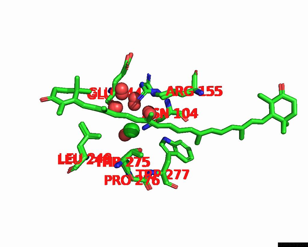
Mono view
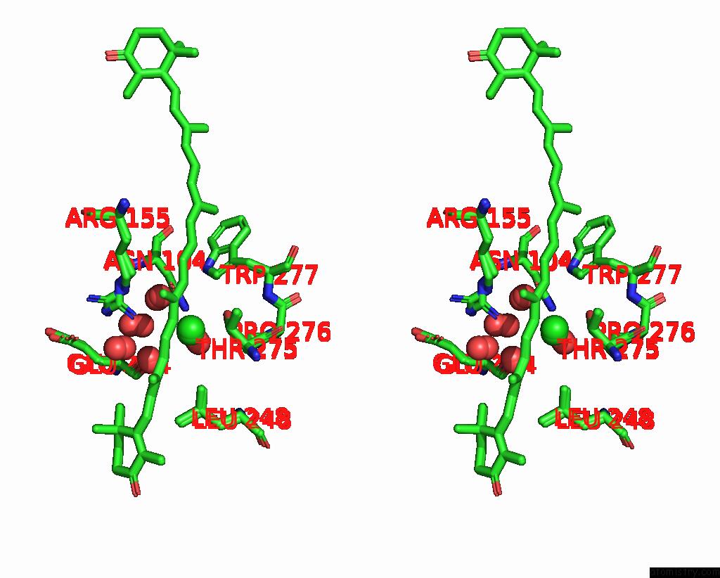
Stereo pair view
Mono view
Stereo pair view
A full contact list of Chlorine with other atoms in the Cl binding
site number 1 of Structure of Orange Carotenoid Protein with Canthaxanthin Bound After 5 Minutes of Illumination within 5.0Å range:
|
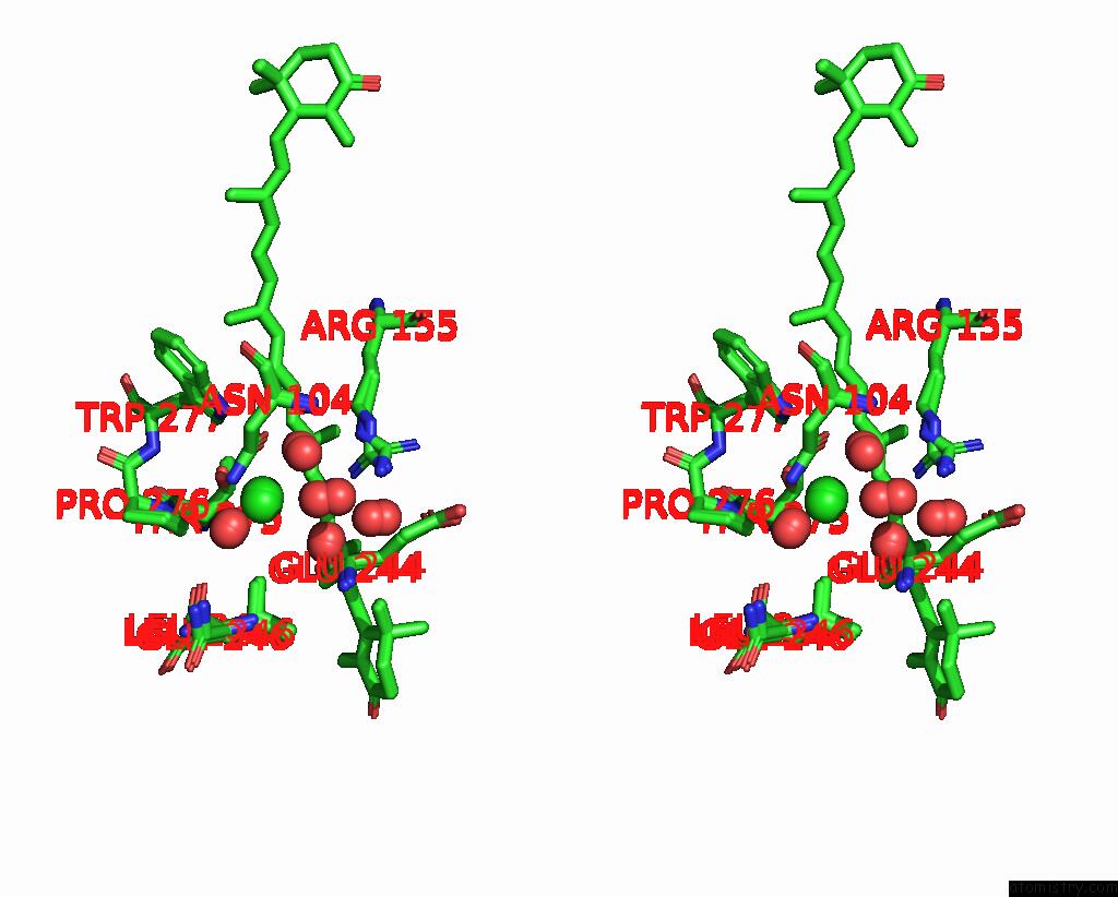
Chlorine binding site 2 out of 2 in 7zsi
Go back to
Chlorine binding site 2 out
of 2 in the Structure of Orange Carotenoid Protein with Canthaxanthin Bound After 5 Minutes of Illumination

Mono view
Stereo pair view
Mono view
Stereo pair view
A full contact list of Chlorine with other atoms in the Cl binding
site number 2 of Structure of Orange Carotenoid Protein with Canthaxanthin Bound After 5 Minutes of Illumination within 5.0Å range:
|
Reference:
V.U.Chukhutsina,
J.M.Baxter,
A.Fadini,
R.M.Morgan,
M.A.Pope,
K.Maghlaoui,
C.M.Orr,
A.Wagner,
J.J.Van Thor.
Light Activation of Orange Carotenoid Protein Reveals Bicycle-Pedal Single-Bond Isomerization. Nat Commun V. 13 6420 2022.
ISSN: ESSN 2041-1723
PubMed: 36307413
DOI: 10.1038/S41467-022-34137-4
Page generated: Tue Jul 30 06:18:32 2024
ISSN: ESSN 2041-1723
PubMed: 36307413
DOI: 10.1038/S41467-022-34137-4
Last articles
Zn in 9JYWZn in 9IR4
Zn in 9IR3
Zn in 9GMX
Zn in 9GMW
Zn in 9JEJ
Zn in 9ERF
Zn in 9ERE
Zn in 9EGV
Zn in 9EGW