Chlorine in PDB 8b6t: X-Ray Structure of the Interface Optimized Haloalkane Dehalogenase HALOTAG7 Fusion to the Green Fluorescent Protein Gfp (CHEMOG5-Tmr) Labeled with A Chloroalkane Tetramethylrhodamine Fluorophore Substrate
Enzymatic activity of X-Ray Structure of the Interface Optimized Haloalkane Dehalogenase HALOTAG7 Fusion to the Green Fluorescent Protein Gfp (CHEMOG5-Tmr) Labeled with A Chloroalkane Tetramethylrhodamine Fluorophore Substrate
All present enzymatic activity of X-Ray Structure of the Interface Optimized Haloalkane Dehalogenase HALOTAG7 Fusion to the Green Fluorescent Protein Gfp (CHEMOG5-Tmr) Labeled with A Chloroalkane Tetramethylrhodamine Fluorophore Substrate:
3.8.1.5;
3.8.1.5;
Protein crystallography data
The structure of X-Ray Structure of the Interface Optimized Haloalkane Dehalogenase HALOTAG7 Fusion to the Green Fluorescent Protein Gfp (CHEMOG5-Tmr) Labeled with A Chloroalkane Tetramethylrhodamine Fluorophore Substrate, PDB code: 8b6t
was solved by
M.Tarnawski,
L.Hellweg,
J.Hiblot,
with X-Ray Crystallography technique. A brief refinement statistics is given in the table below:
| Resolution Low / High (Å) | 46.18 / 2.00 |
| Space group | P 1 21 1 |
| Cell size a, b, c (Å), α, β, γ (°) | 46.6, 64.04, 172.95, 90, 97.67, 90 |
| R / Rfree (%) | 22.1 / 24.5 |
Chlorine Binding Sites:
The binding sites of Chlorine atom in the X-Ray Structure of the Interface Optimized Haloalkane Dehalogenase HALOTAG7 Fusion to the Green Fluorescent Protein Gfp (CHEMOG5-Tmr) Labeled with A Chloroalkane Tetramethylrhodamine Fluorophore Substrate
(pdb code 8b6t). This binding sites where shown within
5.0 Angstroms radius around Chlorine atom.
In total 2 binding sites of Chlorine where determined in the X-Ray Structure of the Interface Optimized Haloalkane Dehalogenase HALOTAG7 Fusion to the Green Fluorescent Protein Gfp (CHEMOG5-Tmr) Labeled with A Chloroalkane Tetramethylrhodamine Fluorophore Substrate, PDB code: 8b6t:
Jump to Chlorine binding site number: 1; 2;
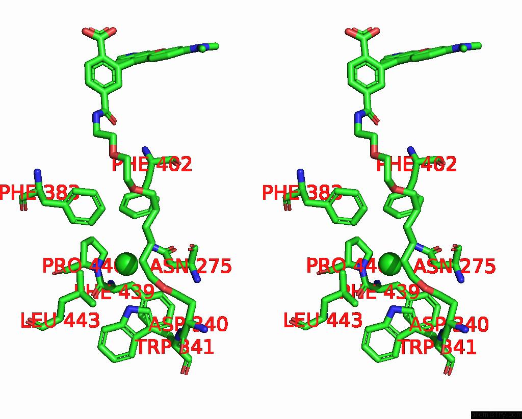
Chlorine binding site 1 out of 2 in 8b6t
Go back to
Chlorine binding site 1 out
of 2 in the X-Ray Structure of the Interface Optimized Haloalkane Dehalogenase HALOTAG7 Fusion to the Green Fluorescent Protein Gfp (CHEMOG5-Tmr) Labeled with A Chloroalkane Tetramethylrhodamine Fluorophore Substrate
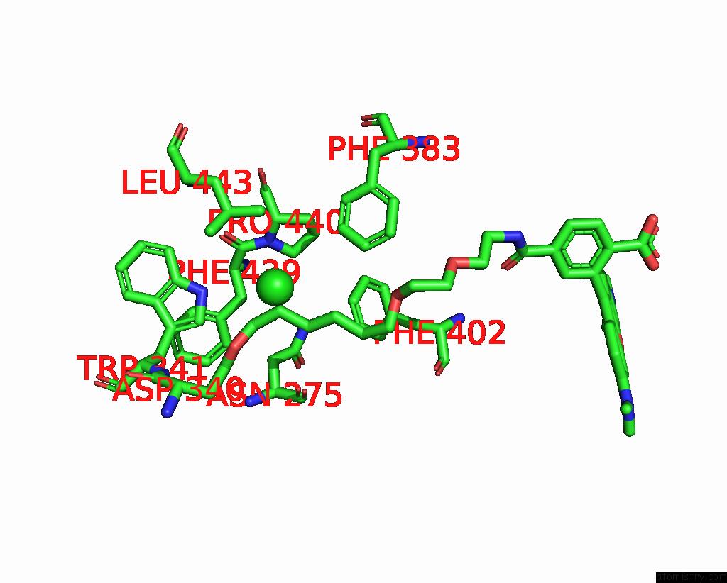
Mono view
Stereo pair view
Mono view
Stereo pair view
A full contact list of Chlorine with other atoms in the Cl binding
site number 1 of X-Ray Structure of the Interface Optimized Haloalkane Dehalogenase HALOTAG7 Fusion to the Green Fluorescent Protein Gfp (CHEMOG5-Tmr) Labeled with A Chloroalkane Tetramethylrhodamine Fluorophore Substrate within 5.0Å range:
|
Chlorine binding site 2 out of 2 in 8b6t
Go back to
Chlorine binding site 2 out
of 2 in the X-Ray Structure of the Interface Optimized Haloalkane Dehalogenase HALOTAG7 Fusion to the Green Fluorescent Protein Gfp (CHEMOG5-Tmr) Labeled with A Chloroalkane Tetramethylrhodamine Fluorophore Substrate

Mono view
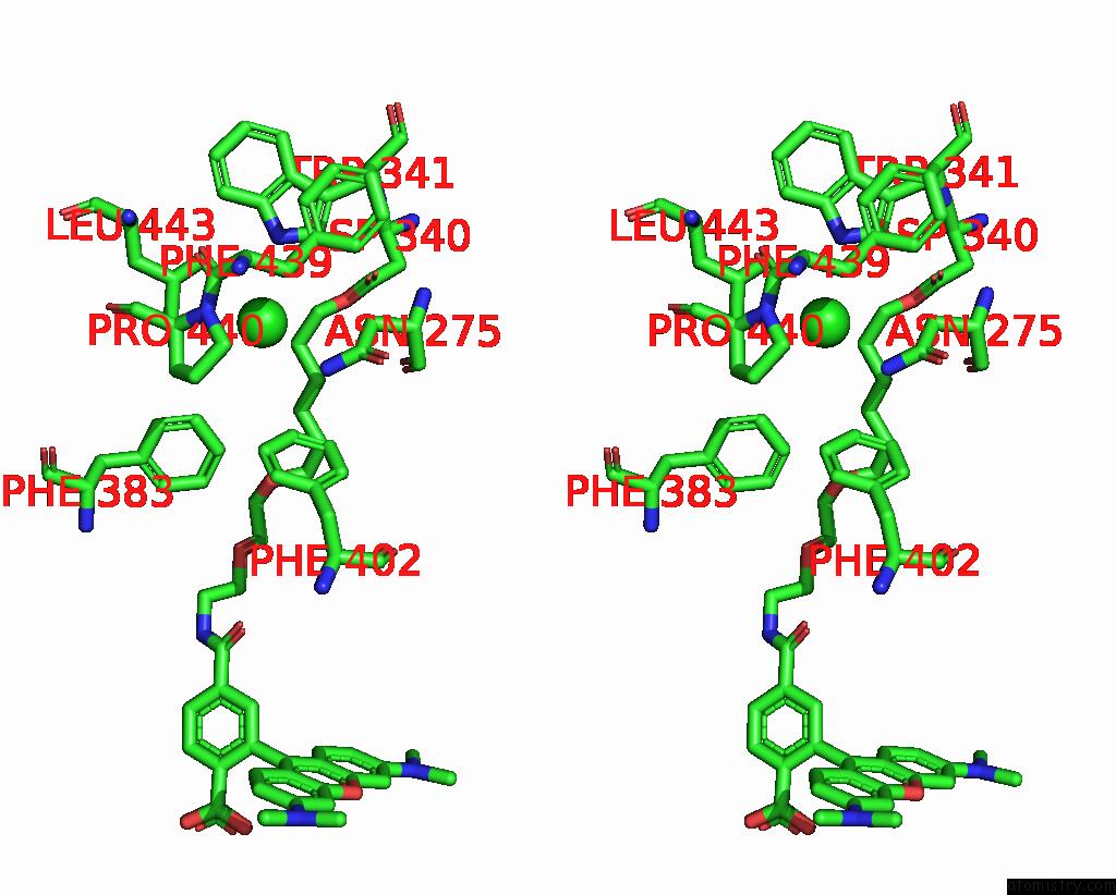
Stereo pair view
Mono view
Stereo pair view
A full contact list of Chlorine with other atoms in the Cl binding
site number 2 of X-Ray Structure of the Interface Optimized Haloalkane Dehalogenase HALOTAG7 Fusion to the Green Fluorescent Protein Gfp (CHEMOG5-Tmr) Labeled with A Chloroalkane Tetramethylrhodamine Fluorophore Substrate within 5.0Å range:
|
Reference:
L.Hellweg,
A.Edenhofer,
L.Barck,
M.C.Huppertz,
M.S.Frei,
M.Tarnawski,
A.Bergner,
B.Koch,
K.Johnsson,
J.Hiblot.
A General Method For the Development of Multicolor Biosensors with Large Dynamic Ranges. Nat.Chem.Biol. 2023.
ISSN: ESSN 1552-4469
PubMed: 37291200
DOI: 10.1038/S41589-023-01350-1
Page generated: Tue Jul 30 07:02:03 2024
ISSN: ESSN 1552-4469
PubMed: 37291200
DOI: 10.1038/S41589-023-01350-1
Last articles
Zn in 9MJ5Zn in 9HNW
Zn in 9G0L
Zn in 9FNE
Zn in 9DZN
Zn in 9E0I
Zn in 9D32
Zn in 9DAK
Zn in 8ZXC
Zn in 8ZUF