Chlorine in PDB 8bvp: Crystal Structure of An N-Terminal Fragment of the Effector Protein LPG2504 (Sidi) From Legionella Pneumophila
Protein crystallography data
The structure of Crystal Structure of An N-Terminal Fragment of the Effector Protein LPG2504 (Sidi) From Legionella Pneumophila, PDB code: 8bvp
was solved by
D.A.Machtens,
J.M.Willerding,
S.Eschenburg,
T.F.Reubold,
with X-Ray Crystallography technique. A brief refinement statistics is given in the table below:
| Resolution Low / High (Å) | 36.12 / 2.10 |
| Space group | C 1 2 1 |
| Cell size a, b, c (Å), α, β, γ (°) | 108.7, 61.03, 113.49, 90, 107.29, 90 |
| R / Rfree (%) | 20.7 / 25.4 |
Chlorine Binding Sites:
The binding sites of Chlorine atom in the Crystal Structure of An N-Terminal Fragment of the Effector Protein LPG2504 (Sidi) From Legionella Pneumophila
(pdb code 8bvp). This binding sites where shown within
5.0 Angstroms radius around Chlorine atom.
In total only one binding site of Chlorine was determined in the Crystal Structure of An N-Terminal Fragment of the Effector Protein LPG2504 (Sidi) From Legionella Pneumophila, PDB code: 8bvp:
In total only one binding site of Chlorine was determined in the Crystal Structure of An N-Terminal Fragment of the Effector Protein LPG2504 (Sidi) From Legionella Pneumophila, PDB code: 8bvp:
Chlorine binding site 1 out of 1 in 8bvp
Go back to
Chlorine binding site 1 out
of 1 in the Crystal Structure of An N-Terminal Fragment of the Effector Protein LPG2504 (Sidi) From Legionella Pneumophila

Mono view
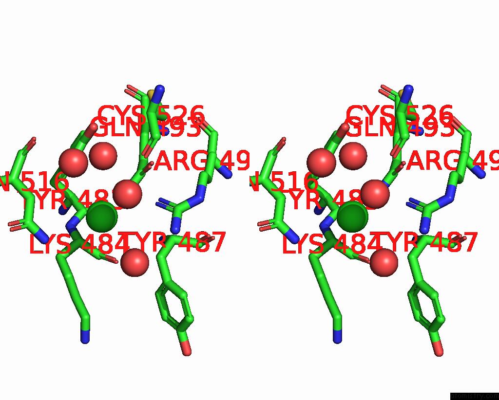
Stereo pair view
Mono view
Stereo pair view
A full contact list of Chlorine with other atoms in the Cl binding
site number 1 of Crystal Structure of An N-Terminal Fragment of the Effector Protein LPG2504 (Sidi) From Legionella Pneumophila within 5.0Å range:
|
Reference:
D.A.Machtens,
J.M.Willerding,
S.Eschenburg,
T.F.Reubold.
Crystal Structure of the N-Terminal Domain of the Effector Protein Sidi of Legionella Pneumophila Reveals A Glucosyl Transferase Domain. Biochem.Biophys.Res.Commun. V. 661 50 2023.
ISSN: ESSN 1090-2104
PubMed: 37087798
DOI: 10.1016/J.BBRC.2023.04.029
Page generated: Tue Jul 30 07:28:57 2024
ISSN: ESSN 1090-2104
PubMed: 37087798
DOI: 10.1016/J.BBRC.2023.04.029
Last articles
Zn in 9JPJZn in 9JP7
Zn in 9JPK
Zn in 9JPL
Zn in 9GN6
Zn in 9GN7
Zn in 9GKU
Zn in 9GKW
Zn in 9GKX
Zn in 9GL0