Chlorine in PDB 8d3g: Crystal Structure of Human Apoptosis-Inducing Factor (Aif) W196A Mutant Complexed with 6-Chloroquinolin-4-Amine
Protein crystallography data
The structure of Crystal Structure of Human Apoptosis-Inducing Factor (Aif) W196A Mutant Complexed with 6-Chloroquinolin-4-Amine, PDB code: 8d3g
was solved by
C.A.Brosey,
J.A.Tainer,
with X-Ray Crystallography technique. A brief refinement statistics is given in the table below:
Resolution Low / High (Å)
|
29.33 /
2.58
|
Space group
|
P 21 21 21
|
Cell size a, b, c (Å), α, β, γ (°)
|
88.296,
110.969,
114.773,
90,
90,
90
|
R / Rfree (%)
|
19.4 /
24.2
|
Chlorine Binding Sites:
The binding sites of Chlorine atom in the Crystal Structure of Human Apoptosis-Inducing Factor (Aif) W196A Mutant Complexed with 6-Chloroquinolin-4-Amine
(pdb code 8d3g). This binding sites where shown within
5.0 Angstroms radius around Chlorine atom.
In total 2 binding sites of Chlorine where determined in the
Crystal Structure of Human Apoptosis-Inducing Factor (Aif) W196A Mutant Complexed with 6-Chloroquinolin-4-Amine, PDB code: 8d3g:
Jump to Chlorine binding site number:
1;
2;
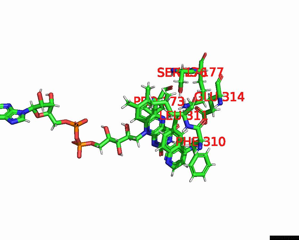
Chlorine binding site 1 out
of 2 in 8d3g
Go back to
Chlorine Binding Sites List in 8d3g
Chlorine binding site 1 out
of 2 in the Crystal Structure of Human Apoptosis-Inducing Factor (Aif) W196A Mutant Complexed with 6-Chloroquinolin-4-Amine
Mono view
Stereo pair view
|
A full contact list of Chlorine with other atoms in the Cl binding
site number 1 of Crystal Structure of Human Apoptosis-Inducing Factor (Aif) W196A Mutant Complexed with 6-Chloroquinolin-4-Amine within 5.0Å range:
probe
|
atom
|
residue
|
distance (Å)
|
B
|
Occ
|
A:Cl702
b:66.6
occ:1.00
|
CL10
|
A:QD1702
|
0.0
|
66.6
|
1.0
|
C09
|
A:QD1702
|
1.8
|
51.0
|
1.0
|
HG
|
A:SER176
|
2.5
|
62.8
|
1.0
|
C11
|
A:QD1702
|
2.8
|
51.8
|
1.0
|
C08
|
A:QD1702
|
2.8
|
45.0
|
1.0
|
HA
|
A:LEU311
|
2.8
|
56.6
|
1.0
|
H111
|
A:QD1702
|
2.9
|
62.1
|
1.0
|
H081
|
A:QD1702
|
2.9
|
54.0
|
1.0
|
OE1
|
A:GLU314
|
3.0
|
58.7
|
1.0
|
HD13
|
A:LEU311
|
3.1
|
51.4
|
1.0
|
H6
|
A:FAD701
|
3.2
|
51.3
|
1.0
|
C6
|
A:FAD701
|
3.3
|
42.8
|
1.0
|
N5
|
A:FAD701
|
3.3
|
43.6
|
1.0
|
C5X
|
A:FAD701
|
3.3
|
43.9
|
1.0
|
OG
|
A:SER176
|
3.4
|
52.3
|
1.0
|
HB3
|
A:PHE310
|
3.5
|
64.9
|
1.0
|
HB2
|
A:LEU311
|
3.6
|
50.9
|
1.0
|
CA
|
A:LEU311
|
3.6
|
47.2
|
1.0
|
CD
|
A:GLU314
|
3.6
|
56.9
|
1.0
|
N
|
A:LEU311
|
3.8
|
43.9
|
1.0
|
HD12
|
A:LEU311
|
3.8
|
51.4
|
1.0
|
HZ2
|
A:LYS177
|
3.8
|
60.1
|
1.0
|
HB2
|
A:GLU314
|
3.8
|
54.0
|
1.0
|
CD1
|
A:LEU311
|
3.8
|
42.9
|
1.0
|
CB
|
A:LEU311
|
4.0
|
42.5
|
1.0
|
C
|
A:PHE310
|
4.0
|
49.1
|
1.0
|
C4X
|
A:FAD701
|
4.1
|
44.6
|
1.0
|
C7
|
A:FAD701
|
4.1
|
44.8
|
1.0
|
C12
|
A:QD1702
|
4.1
|
48.3
|
1.0
|
HZ3
|
A:LYS177
|
4.1
|
60.1
|
1.0
|
C07
|
A:QD1702
|
4.1
|
48.6
|
1.0
|
H
|
A:LEU311
|
4.1
|
52.6
|
1.0
|
OE2
|
A:GLU314
|
4.1
|
56.6
|
1.0
|
O
|
A:PHE310
|
4.2
|
47.5
|
1.0
|
HB2
|
A:PHE310
|
4.2
|
64.9
|
1.0
|
C9A
|
A:FAD701
|
4.2
|
42.8
|
1.0
|
HB3
|
A:SER176
|
4.2
|
64.6
|
1.0
|
CB
|
A:SER176
|
4.2
|
53.9
|
1.0
|
CB
|
A:PHE310
|
4.3
|
54.1
|
1.0
|
HZ1
|
A:LYS177
|
4.3
|
60.1
|
1.0
|
NZ
|
A:LYS177
|
4.3
|
50.1
|
1.0
|
HB2
|
A:SER176
|
4.3
|
64.6
|
1.0
|
HM73
|
A:FAD701
|
4.5
|
55.2
|
1.0
|
CG
|
A:GLU314
|
4.5
|
48.8
|
1.0
|
HG3
|
A:GLU314
|
4.5
|
58.5
|
1.0
|
CB
|
A:GLU314
|
4.5
|
45.0
|
1.0
|
CG
|
A:LEU311
|
4.6
|
46.4
|
1.0
|
HD11
|
A:LEU311
|
4.6
|
51.4
|
1.0
|
C06
|
A:QD1702
|
4.6
|
45.6
|
1.0
|
C4
|
A:FAD701
|
4.7
|
47.1
|
1.0
|
O4
|
A:FAD701
|
4.7
|
51.8
|
1.0
|
HB3
|
A:GLU314
|
4.8
|
54.0
|
1.0
|
C7M
|
A:FAD701
|
4.8
|
46.0
|
1.0
|
CA
|
A:PHE310
|
4.8
|
51.8
|
1.0
|
C
|
A:LEU311
|
4.8
|
46.6
|
1.0
|
C8
|
A:FAD701
|
4.8
|
42.8
|
1.0
|
C10
|
A:FAD701
|
4.9
|
44.0
|
1.0
|
HA
|
A:PRO173
|
4.9
|
52.1
|
1.0
|
N10
|
A:FAD701
|
4.9
|
43.1
|
1.0
|
HB3
|
A:LEU311
|
4.9
|
50.9
|
1.0
|
C9
|
A:FAD701
|
4.9
|
38.9
|
1.0
|
H071
|
A:QD1702
|
4.9
|
58.2
|
1.0
|
|
Chlorine binding site 2 out
of 2 in 8d3g
Go back to
Chlorine Binding Sites List in 8d3g
Chlorine binding site 2 out
of 2 in the Crystal Structure of Human Apoptosis-Inducing Factor (Aif) W196A Mutant Complexed with 6-Chloroquinolin-4-Amine
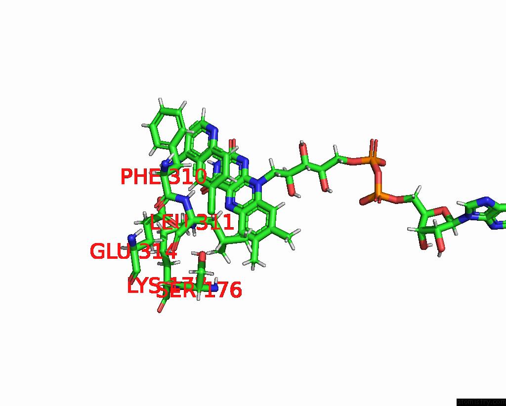
Mono view
Stereo pair view
|
A full contact list of Chlorine with other atoms in the Cl binding
site number 2 of Crystal Structure of Human Apoptosis-Inducing Factor (Aif) W196A Mutant Complexed with 6-Chloroquinolin-4-Amine within 5.0Å range:
probe
|
atom
|
residue
|
distance (Å)
|
B
|
Occ
|
B:Cl702
b:81.7
occ:1.00
|
CL10
|
B:QD1702
|
0.0
|
81.7
|
1.0
|
C09
|
B:QD1702
|
1.8
|
60.7
|
1.0
|
HA
|
B:LEU311
|
2.7
|
64.0
|
1.0
|
C11
|
B:QD1702
|
2.8
|
61.1
|
1.0
|
C08
|
B:QD1702
|
2.8
|
56.1
|
1.0
|
HD13
|
B:LEU311
|
2.8
|
59.3
|
1.0
|
H111
|
B:QD1702
|
2.8
|
73.3
|
1.0
|
H081
|
B:QD1702
|
2.9
|
67.3
|
1.0
|
HB2
|
B:LEU311
|
3.0
|
55.0
|
1.0
|
C5X
|
B:FAD701
|
3.3
|
49.6
|
1.0
|
C6
|
B:FAD701
|
3.3
|
49.8
|
1.0
|
H6
|
B:FAD701
|
3.3
|
59.8
|
1.0
|
N5
|
B:FAD701
|
3.3
|
52.9
|
1.0
|
CA
|
B:LEU311
|
3.4
|
53.4
|
1.0
|
OE1
|
B:GLU314
|
3.4
|
64.4
|
1.0
|
CD1
|
B:LEU311
|
3.6
|
49.4
|
1.0
|
HD12
|
B:LEU311
|
3.6
|
59.3
|
1.0
|
CB
|
B:LEU311
|
3.6
|
45.8
|
1.0
|
HB3
|
B:PHE310
|
3.6
|
82.5
|
1.0
|
N
|
B:LEU311
|
3.6
|
55.0
|
1.0
|
OG
|
B:SER176
|
3.7
|
58.0
|
1.0
|
H
|
B:LEU311
|
3.8
|
66.0
|
1.0
|
C7
|
B:FAD701
|
4.0
|
54.6
|
1.0
|
HB2
|
B:PHE310
|
4.0
|
82.5
|
1.0
|
C9A
|
B:FAD701
|
4.0
|
52.2
|
1.0
|
CD
|
B:GLU314
|
4.0
|
68.9
|
1.0
|
HG
|
B:SER176
|
4.0
|
69.6
|
1.0
|
C
|
B:PHE310
|
4.1
|
62.7
|
1.0
|
C4X
|
B:FAD701
|
4.1
|
53.3
|
1.0
|
HZ3
|
B:LYS177
|
4.1
|
71.4
|
1.0
|
C07
|
B:QD1702
|
4.1
|
60.0
|
1.0
|
C12
|
B:QD1702
|
4.1
|
63.2
|
1.0
|
HB2
|
B:GLU314
|
4.1
|
72.5
|
1.0
|
CG
|
B:LEU311
|
4.2
|
50.8
|
1.0
|
CB
|
B:PHE310
|
4.2
|
68.8
|
1.0
|
HD11
|
B:LEU311
|
4.3
|
59.3
|
1.0
|
O
|
B:PHE310
|
4.4
|
63.5
|
1.0
|
HZ1
|
B:LYS177
|
4.4
|
71.4
|
1.0
|
OE2
|
B:GLU314
|
4.4
|
68.7
|
1.0
|
HD22
|
B:LEU311
|
4.4
|
61.3
|
1.0
|
HM72
|
B:FAD701
|
4.5
|
64.2
|
1.0
|
HB3
|
B:LEU311
|
4.5
|
55.0
|
1.0
|
HZ2
|
B:LYS177
|
4.5
|
71.4
|
1.0
|
NZ
|
B:LYS177
|
4.5
|
59.5
|
1.0
|
HB3
|
B:SER176
|
4.6
|
66.0
|
1.0
|
C8
|
B:FAD701
|
4.6
|
52.3
|
1.0
|
C06
|
B:QD1702
|
4.6
|
64.2
|
1.0
|
C9
|
B:FAD701
|
4.6
|
46.3
|
1.0
|
CB
|
B:SER176
|
4.6
|
55.0
|
1.0
|
N10
|
B:FAD701
|
4.7
|
54.5
|
1.0
|
HB2
|
B:SER176
|
4.7
|
66.0
|
1.0
|
C10
|
B:FAD701
|
4.7
|
52.2
|
1.0
|
C7M
|
B:FAD701
|
4.7
|
53.5
|
1.0
|
C
|
B:LEU311
|
4.7
|
52.8
|
1.0
|
HG3
|
B:GLU314
|
4.8
|
76.0
|
1.0
|
CG
|
B:GLU314
|
4.8
|
63.4
|
1.0
|
C4
|
B:FAD701
|
4.8
|
55.9
|
1.0
|
CA
|
B:PHE310
|
4.8
|
64.2
|
1.0
|
CB
|
B:GLU314
|
4.9
|
60.4
|
1.0
|
H071
|
B:QD1702
|
4.9
|
72.0
|
1.0
|
CD2
|
B:LEU311
|
4.9
|
51.1
|
1.0
|
O4
|
B:FAD701
|
4.9
|
60.5
|
1.0
|
HG
|
B:LEU311
|
5.0
|
61.0
|
1.0
|
|
Reference:
C.A.Brosey,
T.Link,
R.Shen,
D.Moiani,
K.Burnett,
G.Hura,
D.E.Jones,
J.A.Tainer.
Integrating Early Structural Selection Into Chemical Library Screening For Drug Discovery with High-Throughput Small-Angle X-Ray Scattering (Saxs) To Be Published.
Page generated: Tue Jul 30 08:24:47 2024
|