Chlorine in PDB 8d3n: Crystal Structure of Human Apoptosis-Inducing Factor (Aif) Complexed with 7-Chloroquinolin-4-Amine
Protein crystallography data
The structure of Crystal Structure of Human Apoptosis-Inducing Factor (Aif) Complexed with 7-Chloroquinolin-4-Amine, PDB code: 8d3n
was solved by
C.A.Brosey,
J.A.Tainer,
with X-Ray Crystallography technique. A brief refinement statistics is given in the table below:
| Resolution Low / High (Å) | 83.92 / 2.25 |
| Space group | P 21 21 21 |
| Cell size a, b, c (Å), α, β, γ (°) | 91.317, 115.223, 122.479, 90, 90, 90 |
| R / Rfree (%) | 20.7 / 23.7 |
Chlorine Binding Sites:
The binding sites of Chlorine atom in the Crystal Structure of Human Apoptosis-Inducing Factor (Aif) Complexed with 7-Chloroquinolin-4-Amine
(pdb code 8d3n). This binding sites where shown within
5.0 Angstroms radius around Chlorine atom.
In total 2 binding sites of Chlorine where determined in the Crystal Structure of Human Apoptosis-Inducing Factor (Aif) Complexed with 7-Chloroquinolin-4-Amine, PDB code: 8d3n:
Jump to Chlorine binding site number: 1; 2;
In total 2 binding sites of Chlorine where determined in the Crystal Structure of Human Apoptosis-Inducing Factor (Aif) Complexed with 7-Chloroquinolin-4-Amine, PDB code: 8d3n:
Jump to Chlorine binding site number: 1; 2;
Chlorine binding site 1 out of 2 in 8d3n
Go back to
Chlorine binding site 1 out
of 2 in the Crystal Structure of Human Apoptosis-Inducing Factor (Aif) Complexed with 7-Chloroquinolin-4-Amine
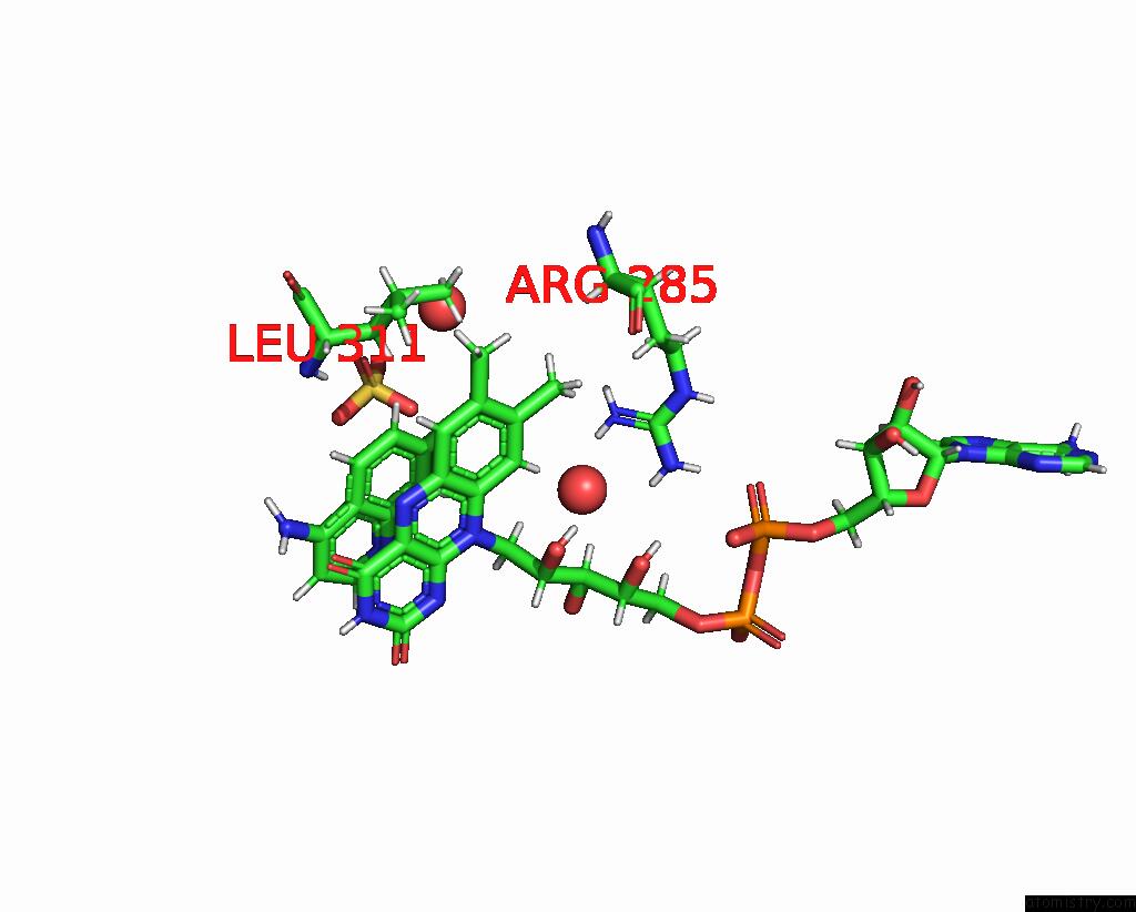
Mono view
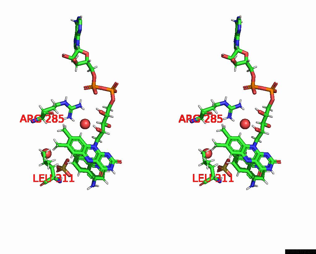
Stereo pair view
Mono view
Stereo pair view
A full contact list of Chlorine with other atoms in the Cl binding
site number 1 of Crystal Structure of Human Apoptosis-Inducing Factor (Aif) Complexed with 7-Chloroquinolin-4-Amine within 5.0Å range:
|
Chlorine binding site 2 out of 2 in 8d3n
Go back to
Chlorine binding site 2 out
of 2 in the Crystal Structure of Human Apoptosis-Inducing Factor (Aif) Complexed with 7-Chloroquinolin-4-Amine
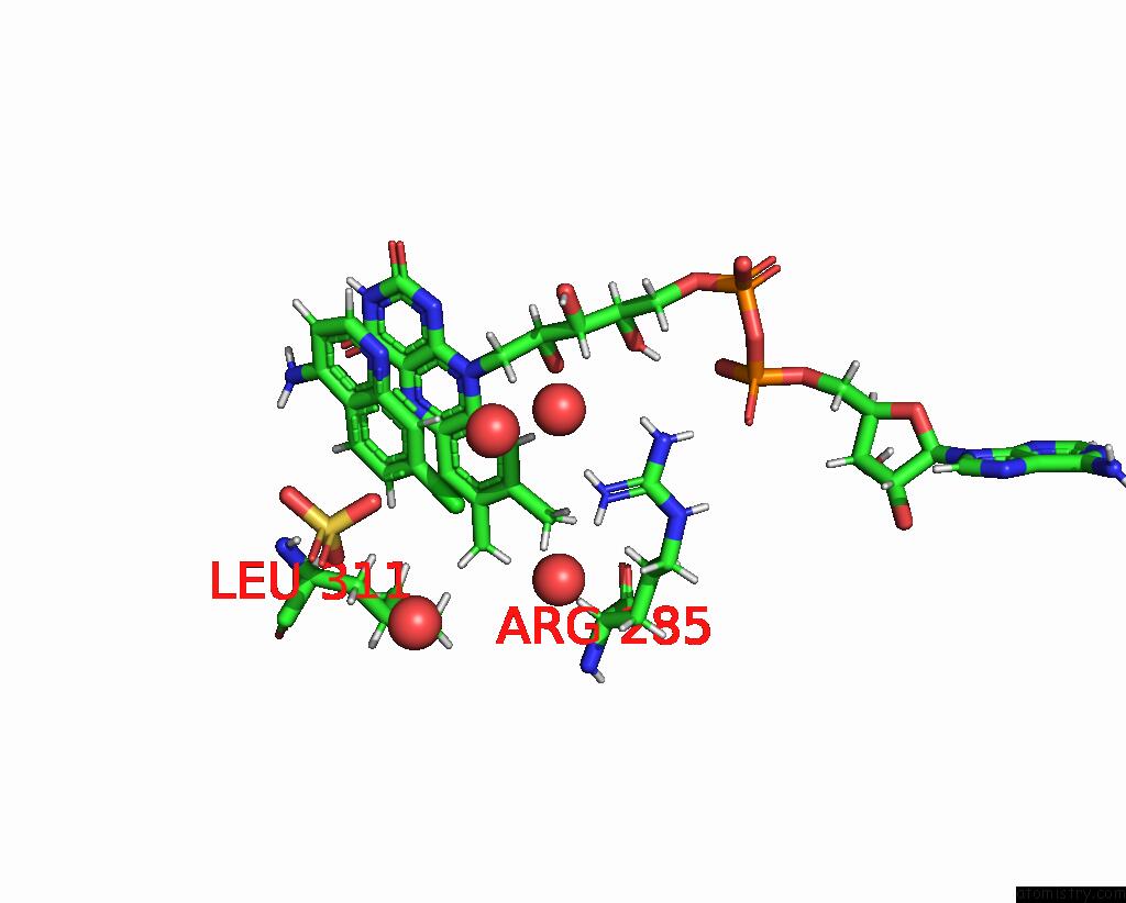
Mono view
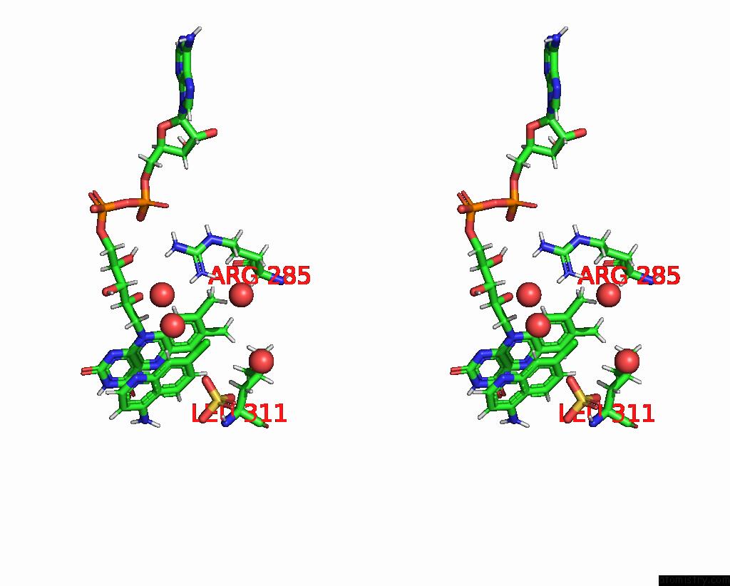
Stereo pair view
Mono view
Stereo pair view
A full contact list of Chlorine with other atoms in the Cl binding
site number 2 of Crystal Structure of Human Apoptosis-Inducing Factor (Aif) Complexed with 7-Chloroquinolin-4-Amine within 5.0Å range:
|
Reference:
C.A.Brosey,
T.Link,
R.Shen,
D.Moiani,
K.Burnett,
G.Hura,
D.E.Jones,
J.A.Tainer.
Integrating Early Structural Selection Into Chemical Library Screening For Drug Discovery with High-Throughput Small-Angle X-Ray Scattering (Saxs) To Be Published.
Page generated: Tue Jul 30 08:25:32 2024
Last articles
Zn in 9JYWZn in 9IR4
Zn in 9IR3
Zn in 9GMX
Zn in 9GMW
Zn in 9JEJ
Zn in 9ERF
Zn in 9ERE
Zn in 9EGV
Zn in 9EGW