Chlorine in PDB 8itz: Crystal Structure of Human Galectin-3 in Complex with Small Molecule Inhibitor
Protein crystallography data
The structure of Crystal Structure of Human Galectin-3 in Complex with Small Molecule Inhibitor, PDB code: 8itz
was solved by
S.Jinal,
K.Amit,
K.Ghosh,
with X-Ray Crystallography technique. A brief refinement statistics is given in the table below:
Resolution Low / High (Å)
|
41.82 /
1.22
|
Space group
|
P 21 21 21
|
Cell size a, b, c (Å), α, β, γ (°)
|
36.51,
57.241,
61.257,
90,
90,
90
|
R / Rfree (%)
|
14.7 /
17.3
|
Other elements in 8itz:
The structure of Crystal Structure of Human Galectin-3 in Complex with Small Molecule Inhibitor also contains other interesting chemical elements:
Chlorine Binding Sites:
The binding sites of Chlorine atom in the Crystal Structure of Human Galectin-3 in Complex with Small Molecule Inhibitor
(pdb code 8itz). This binding sites where shown within
5.0 Angstroms radius around Chlorine atom.
In total 3 binding sites of Chlorine where determined in the
Crystal Structure of Human Galectin-3 in Complex with Small Molecule Inhibitor, PDB code: 8itz:
Jump to Chlorine binding site number:
1;
2;
3;
Chlorine binding site 1 out
of 3 in 8itz
Go back to
Chlorine Binding Sites List in 8itz
Chlorine binding site 1 out
of 3 in the Crystal Structure of Human Galectin-3 in Complex with Small Molecule Inhibitor
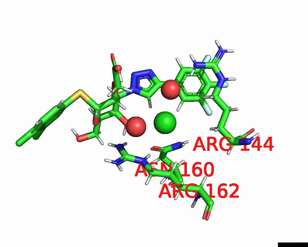
 Mono view
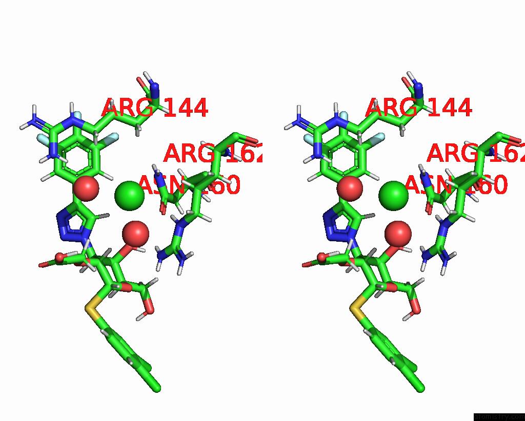
 Stereo pair view
|
A full contact list of Chlorine with other atoms in the Cl binding
site number 1 of Crystal Structure of Human Galectin-3 in Complex with Small Molecule Inhibitor within 5.0Å range:
probe
|
atom
|
residue
|
distance (Å)
|
B
|
Occ
|
A:Cl301
b:18.9
occ:1.00
|
O
|
A:HOH474
|
2.7
|
31.4
|
1.0
|
H15
|
A:QB2302
|
2.8
|
12.5
|
1.0
|
HE
|
A:ARG162
|
3.0
|
10.2
|
1.0
|
HD21
|
A:ASN160
|
3.1
|
9.6
|
1.0
|
O
|
A:HOH546
|
3.3
|
37.8
|
1.0
|
HG2
|
A:ARG162
|
3.3
|
10.3
|
1.0
|
H16
|
A:QB2302
|
3.4
|
12.2
|
1.0
|
OD1
|
A:ASN160
|
3.4
|
10.4
|
1.0
|
NE
|
A:ARG162
|
3.5
|
10.3
|
1.0
|
H2
|
A:QB2302
|
3.5
|
13.2
|
1.0
|
ND2
|
A:ASN160
|
3.7
|
10.0
|
1.0
|
C14
|
A:QB2302
|
3.7
|
11.6
|
1.0
|
HD2
|
A:ARG144
|
3.7
|
15.4
|
1.0
|
H11
|
A:QB2302
|
3.8
|
19.9
|
1.0
|
HB2
|
A:ARG144
|
3.8
|
11.3
|
1.0
|
HH21
|
A:ARG162
|
3.9
|
10.4
|
1.0
|
HD3
|
A:ARG162
|
3.9
|
10.4
|
1.0
|
CG
|
A:ASN160
|
4.0
|
9.5
|
1.0
|
HB3
|
A:ARG144
|
4.0
|
10.8
|
1.0
|
CZ
|
A:ARG162
|
4.1
|
10.8
|
1.0
|
CD
|
A:ARG162
|
4.1
|
10.5
|
1.0
|
CG
|
A:ARG162
|
4.1
|
10.3
|
1.0
|
C17
|
A:QB2302
|
4.1
|
11.9
|
1.0
|
NH2
|
A:ARG162
|
4.2
|
10.6
|
1.0
|
HD3
|
A:ARG144
|
4.2
|
14.9
|
1.0
|
HH21
|
A:ARG144
|
4.3
|
30.2
|
1.0
|
CB
|
A:ARG144
|
4.4
|
11.1
|
1.0
|
O5
|
A:QB2302
|
4.4
|
10.1
|
1.0
|
CD
|
A:ARG144
|
4.4
|
14.7
|
1.0
|
HD22
|
A:ASN160
|
4.4
|
9.6
|
1.0
|
HG3
|
A:ARG162
|
4.5
|
10.4
|
1.0
|
C15
|
A:QB2302
|
4.5
|
14.1
|
1.0
|
C9
|
A:QB2302
|
4.6
|
13.2
|
1.0
|
C16
|
A:QB2302
|
4.6
|
12.6
|
1.0
|
N
|
A:QB2302
|
4.7
|
13.1
|
1.0
|
C10
|
A:QB2302
|
4.7
|
19.4
|
1.0
|
HH22
|
A:ARG162
|
4.8
|
10.1
|
1.0
|
H14
|
A:QB2302
|
4.9
|
10.2
|
1.0
|
C18
|
A:QB2302
|
4.9
|
13.1
|
1.0
|
F
|
A:QB2302
|
5.0
|
14.3
|
1.0
|
NH1
|
A:ARG162
|
5.0
|
11.0
|
1.0
|
CG
|
A:ARG144
|
5.0
|
13.9
|
1.0
|
|
Chlorine binding site 2 out
of 3 in 8itz
Go back to
Chlorine Binding Sites List in 8itz
Chlorine binding site 2 out
of 3 in the Crystal Structure of Human Galectin-3 in Complex with Small Molecule Inhibitor
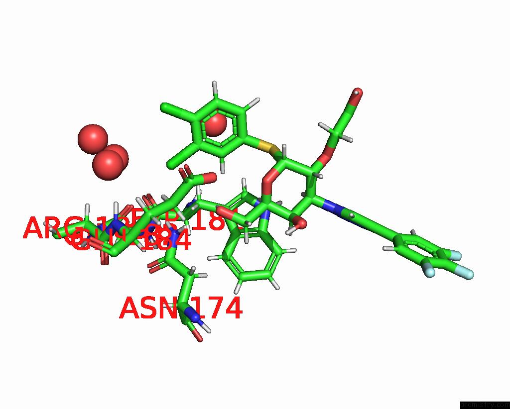
 Mono view
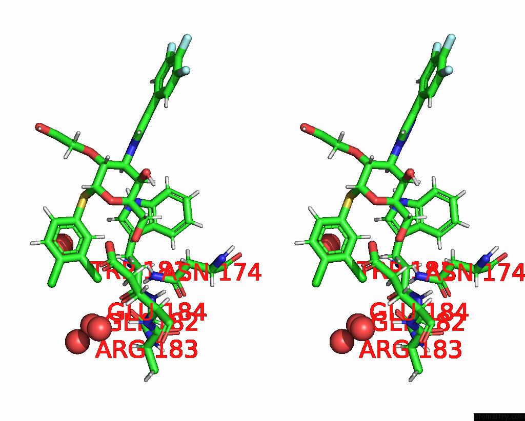
 Stereo pair view
|
A full contact list of Chlorine with other atoms in the Cl binding
site number 2 of Crystal Structure of Human Galectin-3 in Complex with Small Molecule Inhibitor within 5.0Å range:
probe
|
atom
|
residue
|
distance (Å)
|
B
|
Occ
|
A:Cl302
b:15.1
occ:1.00
|
CL1
|
A:QB2302
|
0.0
|
15.1
|
1.0
|
C7
|
A:QB2302
|
1.8
|
13.5
|
1.0
|
C8
|
A:QB2302
|
2.7
|
13.0
|
1.0
|
C6
|
A:QB2302
|
2.7
|
16.9
|
1.0
|
H10
|
A:QB2302
|
2.8
|
13.2
|
1.0
|
HG3
|
A:GLU184
|
3.1
|
12.0
|
1.0
|
HD21
|
A:ASN174
|
3.1
|
9.4
|
1.0
|
CL
|
A:QB2302
|
3.1
|
24.2
|
1.0
|
O
|
A:GLY182
|
3.2
|
11.3
|
1.0
|
H5
|
A:QB2302
|
3.2
|
10.8
|
1.0
|
HB3
|
A:TRP181
|
3.3
|
11.1
|
1.0
|
HD22
|
A:ASN174
|
3.4
|
9.7
|
1.0
|
O
|
A:HOH477
|
3.5
|
27.4
|
1.0
|
HB2
|
A:TRP181
|
3.5
|
11.2
|
1.0
|
ND2
|
A:ASN174
|
3.6
|
10.0
|
1.0
|
O
|
A:QB2302
|
3.7
|
10.8
|
1.0
|
CB
|
A:TRP181
|
3.9
|
11.2
|
1.0
|
OE2
|
A:GLU184
|
3.9
|
12.3
|
1.0
|
CG
|
A:GLU184
|
3.9
|
12.4
|
1.0
|
C3
|
A:QB2302
|
4.0
|
14.0
|
1.0
|
C5
|
A:QB2302
|
4.0
|
18.9
|
1.0
|
CD
|
A:GLU184
|
4.1
|
12.4
|
1.0
|
O
|
A:HOH509
|
4.1
|
18.4
|
1.0
|
HG2
|
A:GLU184
|
4.2
|
11.9
|
1.0
|
O
|
A:HOH572
|
4.3
|
30.0
|
1.0
|
H
|
A:GLU184
|
4.3
|
9.9
|
1.0
|
C
|
A:GLY182
|
4.4
|
11.6
|
1.0
|
C4
|
A:QB2302
|
4.5
|
17.1
|
1.0
|
C
|
A:TRP181
|
4.7
|
12.0
|
1.0
|
O
|
A:HOH593
|
4.7
|
40.6
|
1.0
|
O
|
A:TRP181
|
4.7
|
13.9
|
1.0
|
CG
|
A:TRP181
|
4.8
|
12.3
|
1.0
|
N
|
A:GLU184
|
4.8
|
9.9
|
1.0
|
HA
|
A:ARG183
|
4.9
|
11.4
|
1.0
|
H9
|
A:QB2302
|
4.9
|
17.8
|
1.0
|
N
|
A:GLY182
|
4.9
|
10.4
|
1.0
|
CA
|
A:TRP181
|
4.9
|
11.1
|
1.0
|
CG
|
A:ASN174
|
4.9
|
8.9
|
1.0
|
OE1
|
A:GLU184
|
5.0
|
13.6
|
1.0
|
H
|
A:QB2302
|
5.0
|
11.0
|
1.0
|
C
|
A:QB2302
|
5.0
|
10.1
|
1.0
|
|
Chlorine binding site 3 out
of 3 in 8itz
Go back to
Chlorine Binding Sites List in 8itz
Chlorine binding site 3 out
of 3 in the Crystal Structure of Human Galectin-3 in Complex with Small Molecule Inhibitor
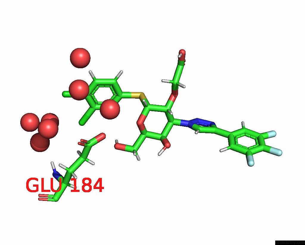
 Mono view
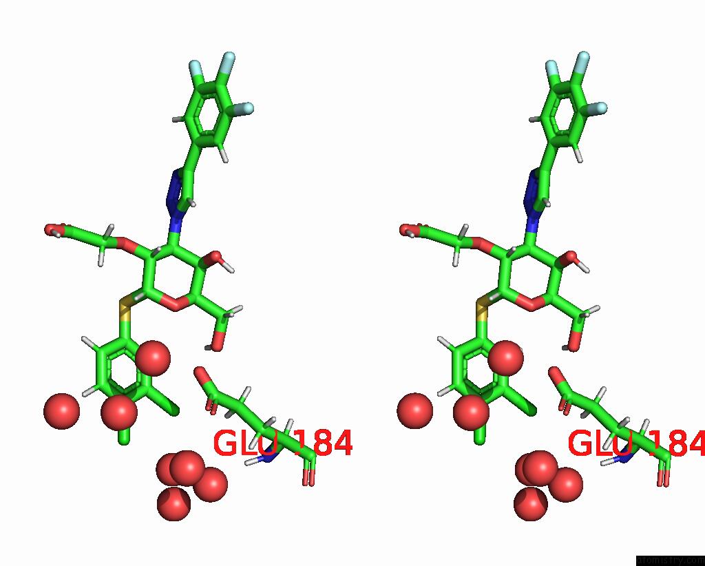
 Stereo pair view
|
A full contact list of Chlorine with other atoms in the Cl binding
site number 3 of Crystal Structure of Human Galectin-3 in Complex with Small Molecule Inhibitor within 5.0Å range:
probe
|
atom
|
residue
|
distance (Å)
|
B
|
Occ
|
A:Cl302
b:24.2
occ:1.00
|
CL
|
A:QB2302
|
0.0
|
24.2
|
1.0
|
C6
|
A:QB2302
|
1.7
|
16.9
|
1.0
|
C5
|
A:QB2302
|
2.7
|
18.9
|
1.0
|
C7
|
A:QB2302
|
2.7
|
13.5
|
1.0
|
H9
|
A:QB2302
|
2.8
|
17.8
|
1.0
|
CL1
|
A:QB2302
|
3.1
|
15.1
|
1.0
|
C4
|
A:QB2302
|
4.0
|
17.1
|
1.0
|
C8
|
A:QB2302
|
4.0
|
13.0
|
1.0
|
O
|
A:HOH426
|
4.0
|
23.6
|
1.0
|
O
|
A:HOH572
|
4.0
|
30.0
|
1.0
|
O
|
A:HOH557
|
4.2
|
22.6
|
1.0
|
O
|
A:HOH597
|
4.2
|
30.0
|
1.0
|
O
|
A:HOH509
|
4.3
|
18.4
|
1.0
|
C3
|
A:QB2302
|
4.5
|
14.0
|
1.0
|
O
|
A:HOH477
|
4.7
|
27.4
|
1.0
|
OE2
|
A:GLU184
|
4.7
|
12.3
|
1.0
|
HG3
|
A:GLU184
|
4.7
|
12.0
|
1.0
|
CD
|
A:GLU184
|
4.8
|
12.4
|
1.0
|
H10
|
A:QB2302
|
4.8
|
13.2
|
1.0
|
H8
|
A:QB2302
|
4.9
|
16.1
|
1.0
|
O
|
A:HOH421
|
4.9
|
13.0
|
1.0
|
OE1
|
A:GLU184
|
5.0
|
13.6
|
1.0
|
|
Reference:
J.J.Swidorski,
B.B.Beno,
C.Liu,
D.Yoon,
K.Ghosh,
H.Sale,
D.Shah,
K.Acharya,
J.Yanchunas,
B.Ellsworth,
D.Cheng,
A.Regueiro-Ren.
Discovery and Exploration of Monosaccharide Linked Dimers to Target Fibrosis To Be Published.
Page generated: Tue Jul 30 10:49:49 2024
|