Chlorine in PDB 8sij: Crystal Structure of F. Varium Tryptophanase
Protein crystallography data
The structure of Crystal Structure of F. Varium Tryptophanase, PDB code: 8sij
was solved by
A.L.Graboski,
M.R.Redinbo,
with X-Ray Crystallography technique. A brief refinement statistics is given in the table below:
| Resolution Low / High (Å) | 48.54 / 2.60 |
| Space group | P 41 21 2 |
| Cell size a, b, c (Å), α, β, γ (°) | 98.589, 98.589, 278.515, 90, 90, 90 |
| R / Rfree (%) | 24 / 26 |
Chlorine Binding Sites:
The binding sites of Chlorine atom in the Crystal Structure of F. Varium Tryptophanase
(pdb code 8sij). This binding sites where shown within
5.0 Angstroms radius around Chlorine atom.
In total 2 binding sites of Chlorine where determined in the Crystal Structure of F. Varium Tryptophanase, PDB code: 8sij:
Jump to Chlorine binding site number: 1; 2;
In total 2 binding sites of Chlorine where determined in the Crystal Structure of F. Varium Tryptophanase, PDB code: 8sij:
Jump to Chlorine binding site number: 1; 2;
Chlorine binding site 1 out of 2 in 8sij
Go back to
Chlorine binding site 1 out
of 2 in the Crystal Structure of F. Varium Tryptophanase

Mono view
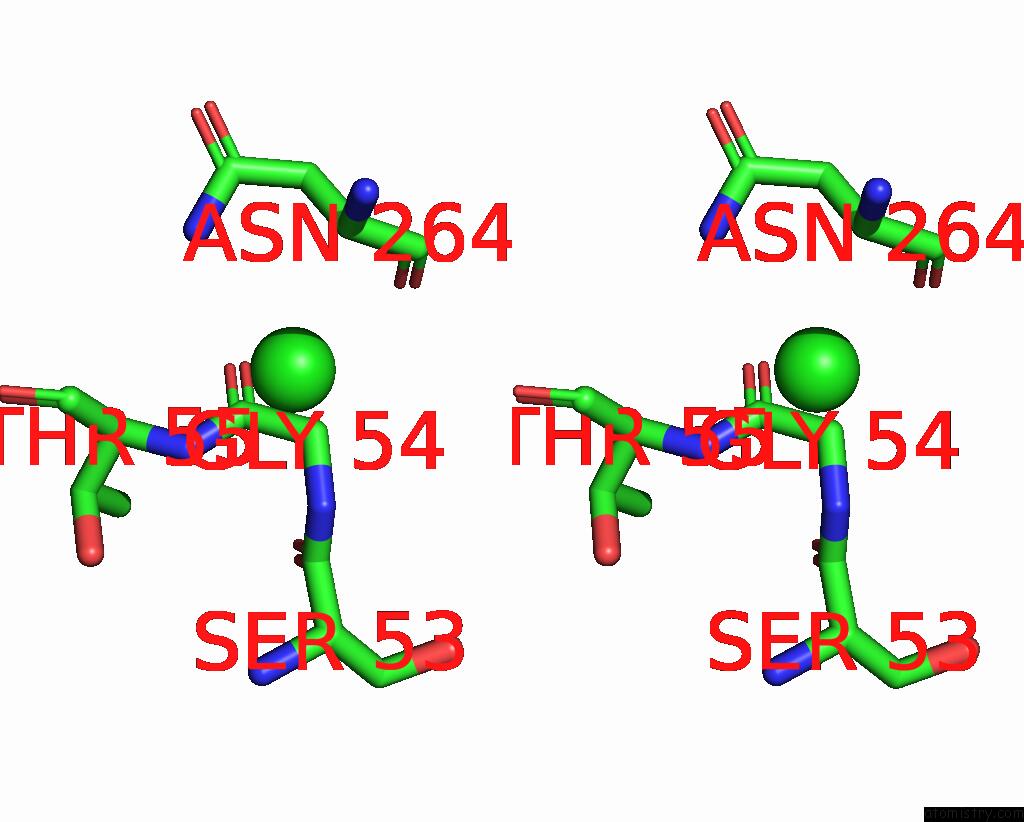
Stereo pair view
Mono view
Stereo pair view
A full contact list of Chlorine with other atoms in the Cl binding
site number 1 of Crystal Structure of F. Varium Tryptophanase within 5.0Å range:
|
Chlorine binding site 2 out of 2 in 8sij
Go back to
Chlorine binding site 2 out
of 2 in the Crystal Structure of F. Varium Tryptophanase
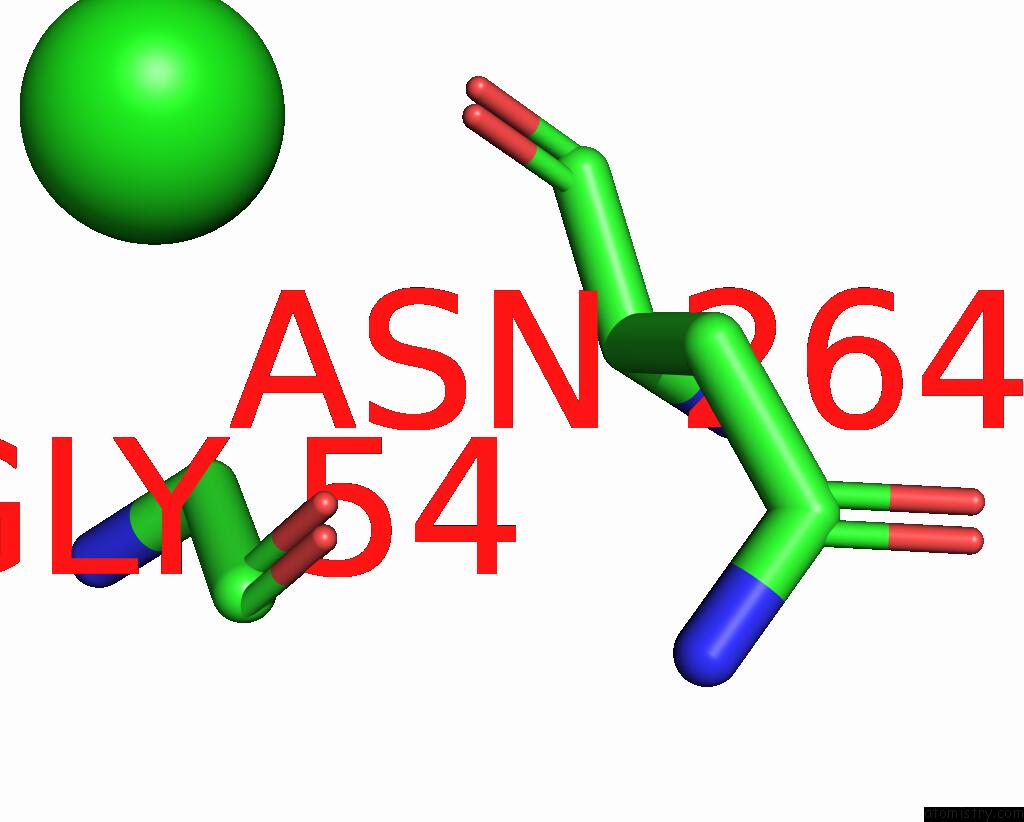
Mono view
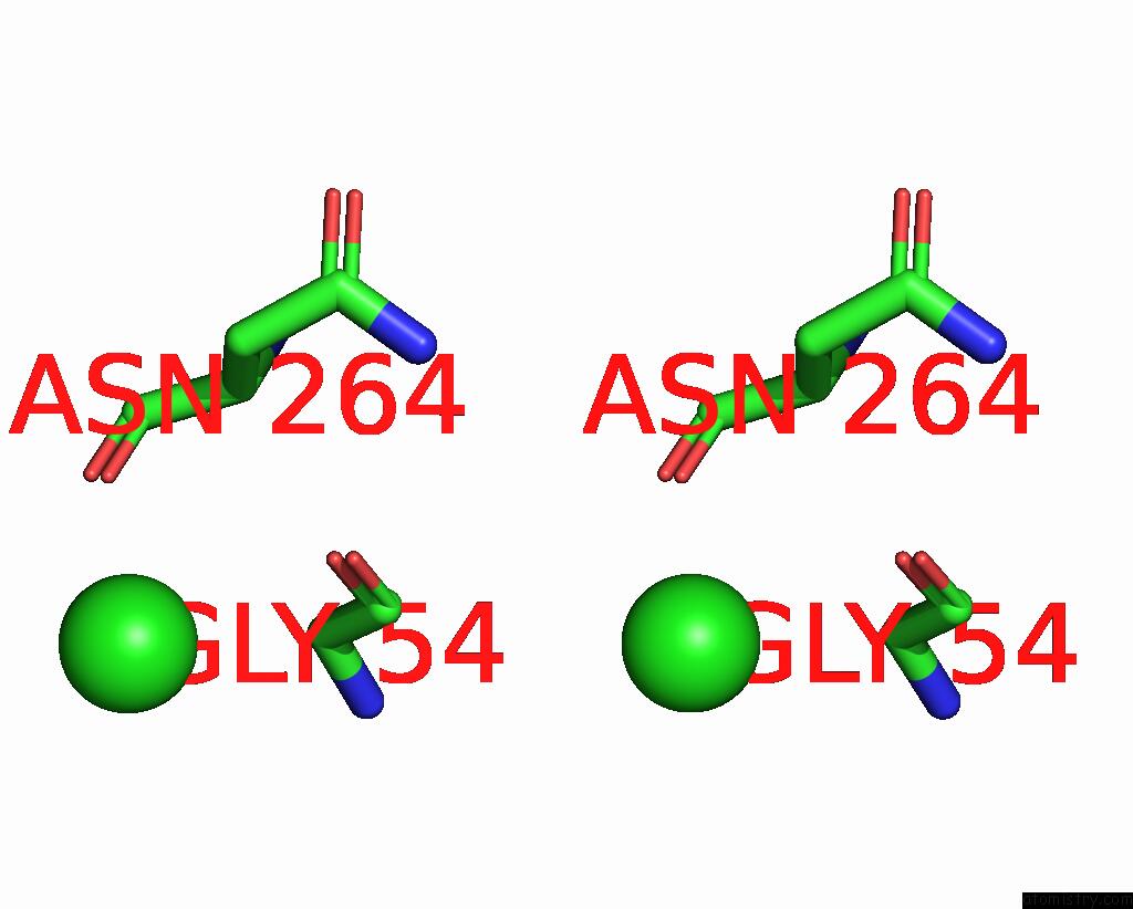
Stereo pair view
Mono view
Stereo pair view
A full contact list of Chlorine with other atoms in the Cl binding
site number 2 of Crystal Structure of F. Varium Tryptophanase within 5.0Å range:
|
Reference:
A.L.Graboski,
M.R.Redinbo.
Mechanism-Based Inhibition of Gut Microbial Tryptophanases Reduces Serum Indoxyl Sulfate Cell Chem Biol 2023.
ISSN: ESSN 2451-9456
DOI: 10.1016/J.CHEMBIOL.2023.07.015
Page generated: Tue Jul 30 12:27:27 2024
ISSN: ESSN 2451-9456
DOI: 10.1016/J.CHEMBIOL.2023.07.015
Last articles
Zn in 9MJ5Zn in 9HNW
Zn in 9G0L
Zn in 9FNE
Zn in 9DZN
Zn in 9E0I
Zn in 9D32
Zn in 9DAK
Zn in 8ZXC
Zn in 8ZUF