Chlorine in PDB 8sxw: X-Ray Crystal Structure of Udp- 2,3-Diacetamido-2,3-Dideoxy-Glucuronic Acid-2-Epimerase From Thermus Thermophilus Strain HB27, D98N Mutation, Apo Structure at pH 6
Protein crystallography data
The structure of X-Ray Crystal Structure of Udp- 2,3-Diacetamido-2,3-Dideoxy-Glucuronic Acid-2-Epimerase From Thermus Thermophilus Strain HB27, D98N Mutation, Apo Structure at pH 6, PDB code: 8sxw
was solved by
C.W.Kroft,
J.B.Thoden,
H.M.Holden,
with X-Ray Crystallography technique. A brief refinement statistics is given in the table below:
| Resolution Low / High (Å) | 41.72 / 1.80 |
| Space group | P 1 |
| Cell size a, b, c (Å), α, β, γ (°) | 64.327, 85.34, 86.535, 108.23, 110.06, 97.1 |
| R / Rfree (%) | 17.9 / 22.4 |
Other elements in 8sxw:
The structure of X-Ray Crystal Structure of Udp- 2,3-Diacetamido-2,3-Dideoxy-Glucuronic Acid-2-Epimerase From Thermus Thermophilus Strain HB27, D98N Mutation, Apo Structure at pH 6 also contains other interesting chemical elements:
| Sodium | (Na) | 1 atom |
Chlorine Binding Sites:
The binding sites of Chlorine atom in the X-Ray Crystal Structure of Udp- 2,3-Diacetamido-2,3-Dideoxy-Glucuronic Acid-2-Epimerase From Thermus Thermophilus Strain HB27, D98N Mutation, Apo Structure at pH 6
(pdb code 8sxw). This binding sites where shown within
5.0 Angstroms radius around Chlorine atom.
In total 2 binding sites of Chlorine where determined in the X-Ray Crystal Structure of Udp- 2,3-Diacetamido-2,3-Dideoxy-Glucuronic Acid-2-Epimerase From Thermus Thermophilus Strain HB27, D98N Mutation, Apo Structure at pH 6, PDB code: 8sxw:
Jump to Chlorine binding site number: 1; 2;
In total 2 binding sites of Chlorine where determined in the X-Ray Crystal Structure of Udp- 2,3-Diacetamido-2,3-Dideoxy-Glucuronic Acid-2-Epimerase From Thermus Thermophilus Strain HB27, D98N Mutation, Apo Structure at pH 6, PDB code: 8sxw:
Jump to Chlorine binding site number: 1; 2;
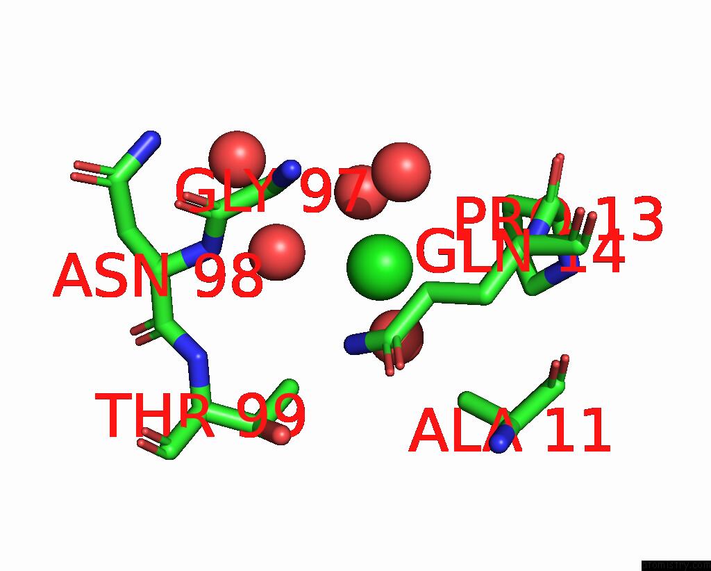
Chlorine binding site 1 out of 2 in 8sxw
Go back to
Chlorine binding site 1 out
of 2 in the X-Ray Crystal Structure of Udp- 2,3-Diacetamido-2,3-Dideoxy-Glucuronic Acid-2-Epimerase From Thermus Thermophilus Strain HB27, D98N Mutation, Apo Structure at pH 6

Mono view

Stereo pair view
Mono view
Stereo pair view
A full contact list of Chlorine with other atoms in the Cl binding
site number 1 of X-Ray Crystal Structure of Udp- 2,3-Diacetamido-2,3-Dideoxy-Glucuronic Acid-2-Epimerase From Thermus Thermophilus Strain HB27, D98N Mutation, Apo Structure at pH 6 within 5.0Å range:
|
Chlorine binding site 2 out of 2 in 8sxw
Go back to
Chlorine binding site 2 out
of 2 in the X-Ray Crystal Structure of Udp- 2,3-Diacetamido-2,3-Dideoxy-Glucuronic Acid-2-Epimerase From Thermus Thermophilus Strain HB27, D98N Mutation, Apo Structure at pH 6
Mono view

Stereo pair view
Mono view
Stereo pair view
A full contact list of Chlorine with other atoms in the Cl binding
site number 2 of X-Ray Crystal Structure of Udp- 2,3-Diacetamido-2,3-Dideoxy-Glucuronic Acid-2-Epimerase From Thermus Thermophilus Strain HB27, D98N Mutation, Apo Structure at pH 6 within 5.0Å range:
|
Reference:
J.B.Thoden,
J.O.Mcknight,
C.W.Kroft,
J.D.T.Jast,
H.M.Holden.
Structural Analysis of A Bacterial Udp-Sugar 2-Epimerase Reveals the Active Site Architecture Before and After Catalysis. J.Biol.Chem. 05200 2023.
ISSN: ESSN 1083-351X
PubMed: 37660908
DOI: 10.1016/J.JBC.2023.105200
Page generated: Tue Jul 30 12:39:47 2024
ISSN: ESSN 1083-351X
PubMed: 37660908
DOI: 10.1016/J.JBC.2023.105200
Last articles
Zn in 9J0NZn in 9J0O
Zn in 9J0P
Zn in 9FJX
Zn in 9EKB
Zn in 9C0F
Zn in 9CAH
Zn in 9CH0
Zn in 9CH3
Zn in 9CH1