Chlorine in PDB 8xnj: Crystal Structure of Trypsin in-Complex with Arginine
Enzymatic activity of Crystal Structure of Trypsin in-Complex with Arginine
All present enzymatic activity of Crystal Structure of Trypsin in-Complex with Arginine:
3.4.21.4;
3.4.21.4;
Protein crystallography data
The structure of Crystal Structure of Trypsin in-Complex with Arginine, PDB code: 8xnj
was solved by
Z.Akbar,
M.S.Ahmad,
with X-Ray Crystallography technique. A brief refinement statistics is given in the table below:
| Resolution Low / High (Å) | 19.81 / 2.40 |
| Space group | P 21 21 21 |
| Cell size a, b, c (Å), α, β, γ (°) | 54.101, 58.152, 66.288, 90, 90, 90 |
| R / Rfree (%) | 19.2 / 27.5 |
Other elements in 8xnj:
The structure of Crystal Structure of Trypsin in-Complex with Arginine also contains other interesting chemical elements:
| Calcium | (Ca) | 1 atom |
Chlorine Binding Sites:
The binding sites of Chlorine atom in the Crystal Structure of Trypsin in-Complex with Arginine
(pdb code 8xnj). This binding sites where shown within
5.0 Angstroms radius around Chlorine atom.
In total 3 binding sites of Chlorine where determined in the Crystal Structure of Trypsin in-Complex with Arginine, PDB code: 8xnj:
Jump to Chlorine binding site number: 1; 2; 3;
In total 3 binding sites of Chlorine where determined in the Crystal Structure of Trypsin in-Complex with Arginine, PDB code: 8xnj:
Jump to Chlorine binding site number: 1; 2; 3;
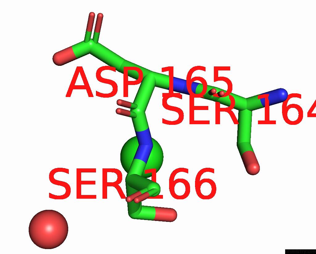
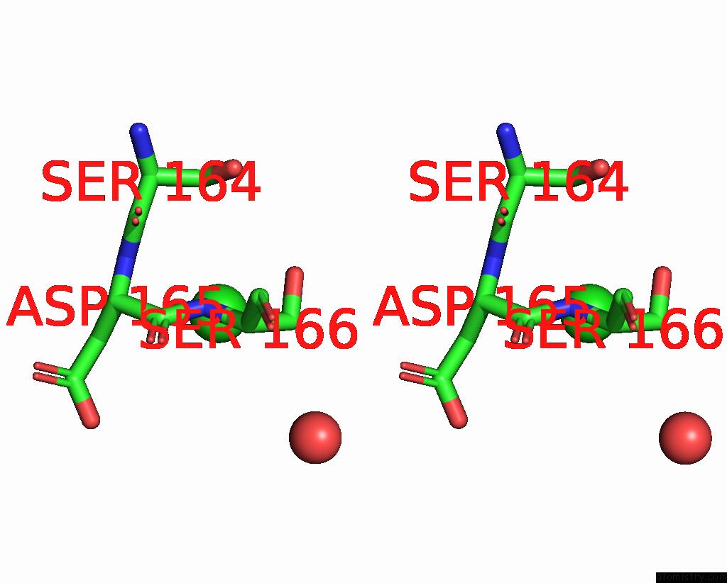
Chlorine binding site 1 out of 3 in 8xnj
Go back to
Chlorine binding site 1 out
of 3 in the Crystal Structure of Trypsin in-Complex with Arginine
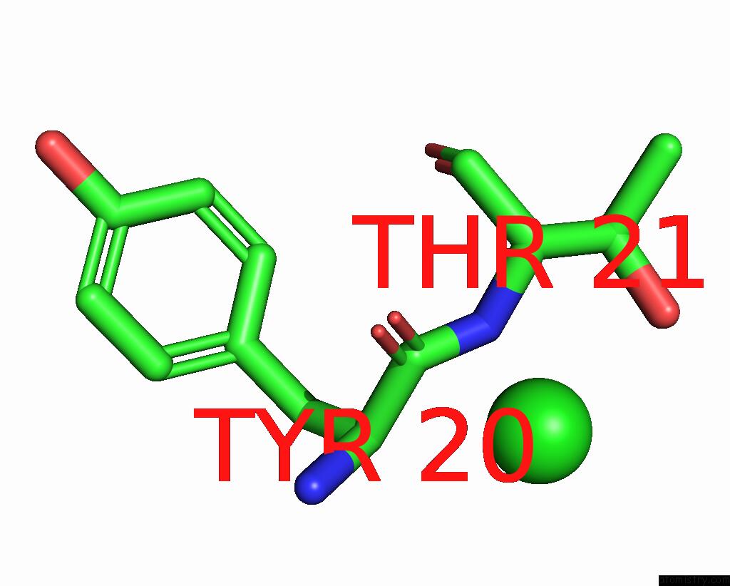
Mono view
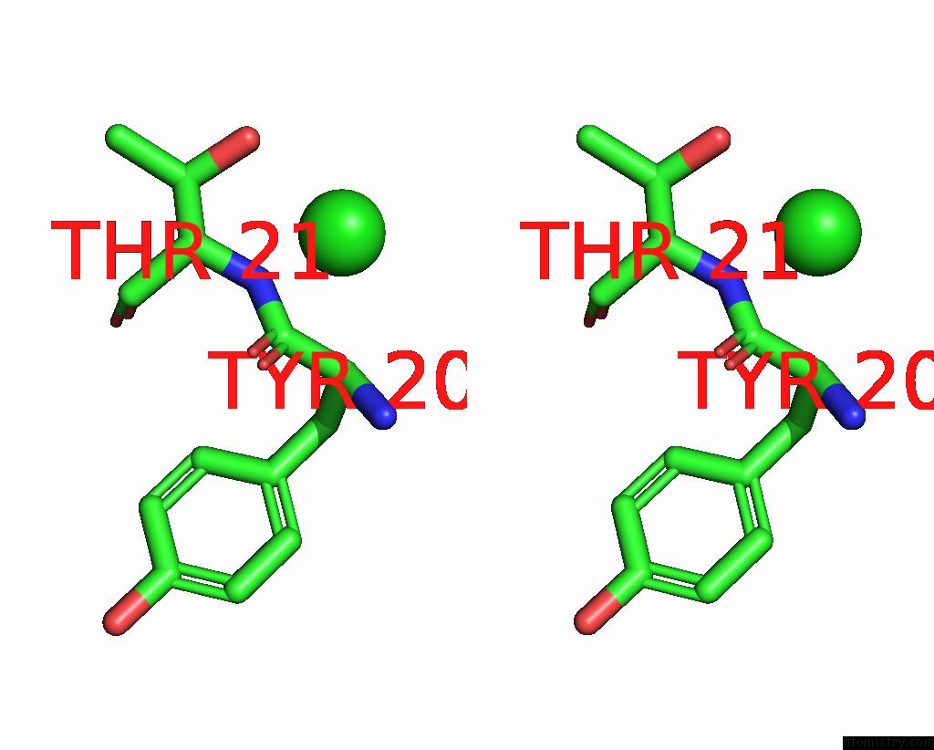
Stereo pair view
Mono view
Stereo pair view
A full contact list of Chlorine with other atoms in the Cl binding
site number 1 of Crystal Structure of Trypsin in-Complex with Arginine within 5.0Å range:
|
Chlorine binding site 2 out of 3 in 8xnj
Go back to
Chlorine binding site 2 out
of 3 in the Crystal Structure of Trypsin in-Complex with Arginine
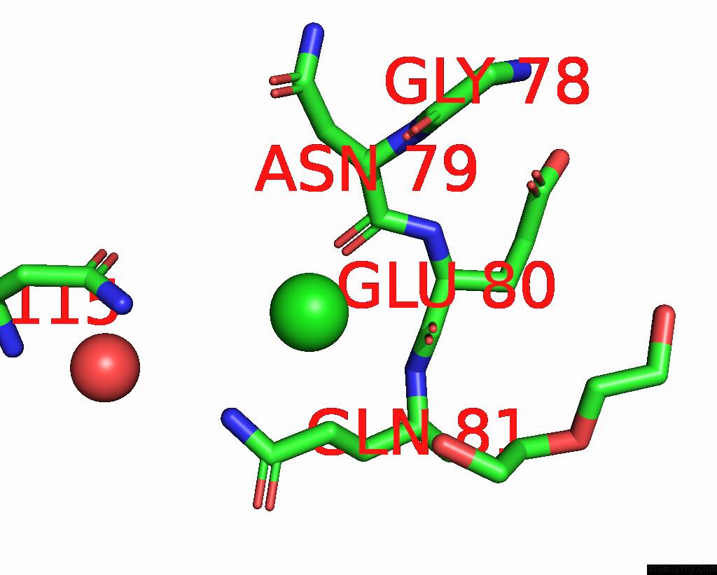
Mono view
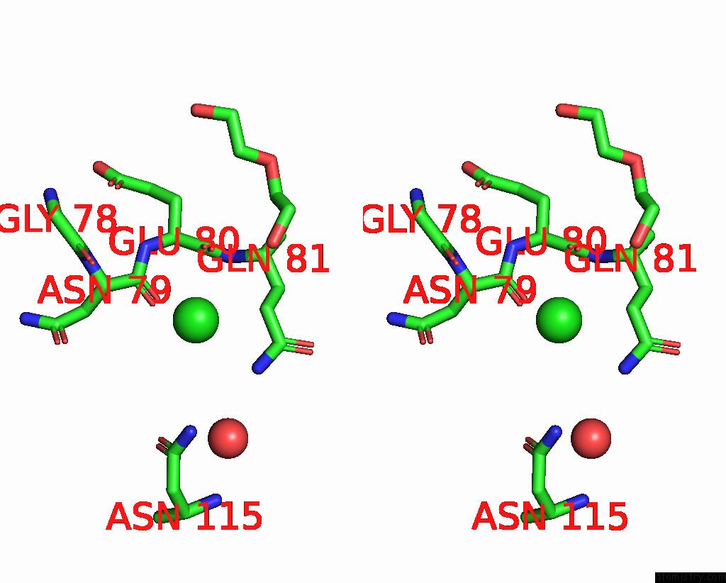
Stereo pair view
Mono view
Stereo pair view
A full contact list of Chlorine with other atoms in the Cl binding
site number 2 of Crystal Structure of Trypsin in-Complex with Arginine within 5.0Å range:
|
Chlorine binding site 3 out of 3 in 8xnj
Go back to
Chlorine binding site 3 out
of 3 in the Crystal Structure of Trypsin in-Complex with Arginine
Mono view
Stereo pair view
Mono view
Stereo pair view
A full contact list of Chlorine with other atoms in the Cl binding
site number 3 of Crystal Structure of Trypsin in-Complex with Arginine within 5.0Å range:
|
Reference:
Z.Akbar,
M.S.Ahmad.
In Vitro , in Silico and Crystallographic-Based Identification of Serine Protease Inhibitors. Nat Prod Res 1 2024.
ISSN: ISSN 1478-6427
PubMed: 39520718
DOI: 10.1080/14786419.2024.2425793
Page generated: Tue Dec 10 19:28:21 2024
ISSN: ISSN 1478-6427
PubMed: 39520718
DOI: 10.1080/14786419.2024.2425793
Last articles
Zn in 9IRQZn in 9IYX
Zn in 9J8P
Zn in 9IUU
Zn in 9GBF
Zn in 9G2V
Zn in 9G2L
Zn in 9G2X
Zn in 9G2Z
Zn in 9G2K