Chlorine in PDB 8zur: Crystal Structure of the F99S/M153T/V163A/T203V/E222Q Variant of Gfp at pH 5.0
Protein crystallography data
The structure of Crystal Structure of the F99S/M153T/V163A/T203V/E222Q Variant of Gfp at pH 5.0, PDB code: 8zur
was solved by
R.Takeda,
K.Takeda,
with X-Ray Crystallography technique. A brief refinement statistics is given in the table below:
| Resolution Low / High (Å) | 41.20 / 1.20 |
| Space group | P 21 21 21 |
| Cell size a, b, c (Å), α, β, γ (°) | 51.5, 57.79, 68.67, 90, 90, 90 |
| R / Rfree (%) | 12.9 / 16.6 |
Chlorine Binding Sites:
The binding sites of Chlorine atom in the Crystal Structure of the F99S/M153T/V163A/T203V/E222Q Variant of Gfp at pH 5.0
(pdb code 8zur). This binding sites where shown within
5.0 Angstroms radius around Chlorine atom.
In total 2 binding sites of Chlorine where determined in the Crystal Structure of the F99S/M153T/V163A/T203V/E222Q Variant of Gfp at pH 5.0, PDB code: 8zur:
Jump to Chlorine binding site number: 1; 2;
In total 2 binding sites of Chlorine where determined in the Crystal Structure of the F99S/M153T/V163A/T203V/E222Q Variant of Gfp at pH 5.0, PDB code: 8zur:
Jump to Chlorine binding site number: 1; 2;
Chlorine binding site 1 out of 2 in 8zur
Go back to
Chlorine binding site 1 out
of 2 in the Crystal Structure of the F99S/M153T/V163A/T203V/E222Q Variant of Gfp at pH 5.0
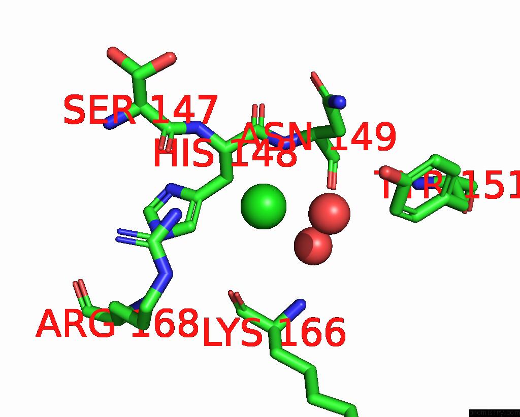
Mono view
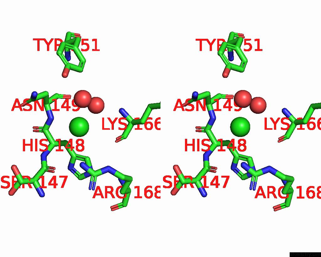
Stereo pair view
Mono view
Stereo pair view
A full contact list of Chlorine with other atoms in the Cl binding
site number 1 of Crystal Structure of the F99S/M153T/V163A/T203V/E222Q Variant of Gfp at pH 5.0 within 5.0Å range:
|
Chlorine binding site 2 out of 2 in 8zur
Go back to
Chlorine binding site 2 out
of 2 in the Crystal Structure of the F99S/M153T/V163A/T203V/E222Q Variant of Gfp at pH 5.0
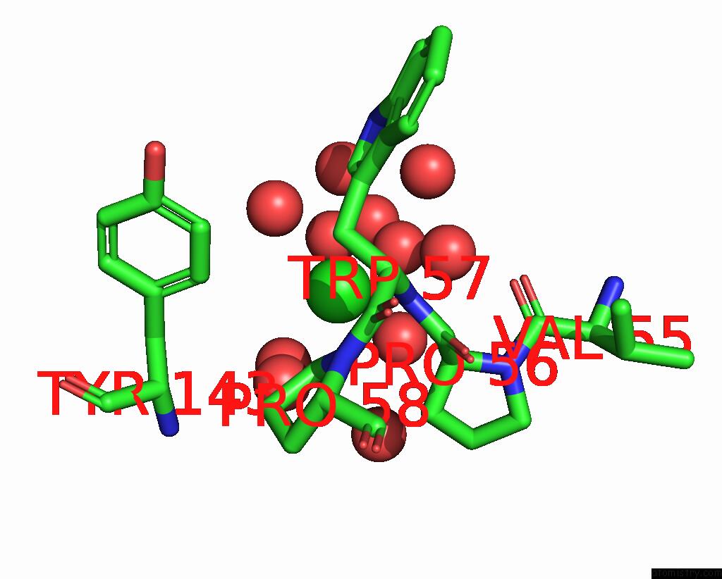
Mono view
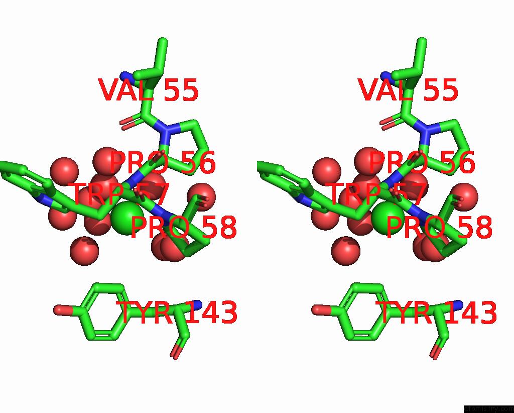
Stereo pair view
Mono view
Stereo pair view
A full contact list of Chlorine with other atoms in the Cl binding
site number 2 of Crystal Structure of the F99S/M153T/V163A/T203V/E222Q Variant of Gfp at pH 5.0 within 5.0Å range:
|
Reference:
R.Takeda,
E.Tsutsumi,
K.Okatsu,
S.Fukai,
K.Takeda.
Structural Characterization of Green Fluorescent Protein in the I-State. Sci Rep V. 14 22832 2024.
ISSN: ESSN 2045-2322
PubMed: 39353998
DOI: 10.1038/S41598-024-73696-Y
Page generated: Wed Nov 13 07:52:36 2024
ISSN: ESSN 2045-2322
PubMed: 39353998
DOI: 10.1038/S41598-024-73696-Y
Last articles
Zn in 9MJ5Zn in 9HNW
Zn in 9G0L
Zn in 9FNE
Zn in 9DZN
Zn in 9E0I
Zn in 9D32
Zn in 9DAK
Zn in 8ZXC
Zn in 8ZUF