Chlorine »
PDB 8ayq-8b7b »
8b6r »
Chlorine in PDB 8b6r: X-Ray Structure of the Haloalkane Dehalogenase HALOTAG7 Labeled with A Chloroalkane CYANINE3 Fluorophore Substrate
Enzymatic activity of X-Ray Structure of the Haloalkane Dehalogenase HALOTAG7 Labeled with A Chloroalkane CYANINE3 Fluorophore Substrate
All present enzymatic activity of X-Ray Structure of the Haloalkane Dehalogenase HALOTAG7 Labeled with A Chloroalkane CYANINE3 Fluorophore Substrate:
3.8.1.5;
3.8.1.5;
Protein crystallography data
The structure of X-Ray Structure of the Haloalkane Dehalogenase HALOTAG7 Labeled with A Chloroalkane CYANINE3 Fluorophore Substrate, PDB code: 8b6r
was solved by
M.Tarnawski,
L.Hellweg,
J.Hiblot,
with X-Ray Crystallography technique. A brief refinement statistics is given in the table below:
| Resolution Low / High (Å) | 41.25 / 1.50 |
| Space group | P 42 21 2 |
| Cell size a, b, c (Å), α, β, γ (°) | 112.56, 112.56, 44.33, 90, 90, 90 |
| R / Rfree (%) | 16.2 / 19.2 |
Other elements in 8b6r:
The structure of X-Ray Structure of the Haloalkane Dehalogenase HALOTAG7 Labeled with A Chloroalkane CYANINE3 Fluorophore Substrate also contains other interesting chemical elements:
| Magnesium | (Mg) | 1 atom |
Chlorine Binding Sites:
The binding sites of Chlorine atom in the X-Ray Structure of the Haloalkane Dehalogenase HALOTAG7 Labeled with A Chloroalkane CYANINE3 Fluorophore Substrate
(pdb code 8b6r). This binding sites where shown within
5.0 Angstroms radius around Chlorine atom.
In total only one binding site of Chlorine was determined in the X-Ray Structure of the Haloalkane Dehalogenase HALOTAG7 Labeled with A Chloroalkane CYANINE3 Fluorophore Substrate, PDB code: 8b6r:
In total only one binding site of Chlorine was determined in the X-Ray Structure of the Haloalkane Dehalogenase HALOTAG7 Labeled with A Chloroalkane CYANINE3 Fluorophore Substrate, PDB code: 8b6r:
Chlorine binding site 1 out of 1 in 8b6r
Go back to
Chlorine binding site 1 out
of 1 in the X-Ray Structure of the Haloalkane Dehalogenase HALOTAG7 Labeled with A Chloroalkane CYANINE3 Fluorophore Substrate
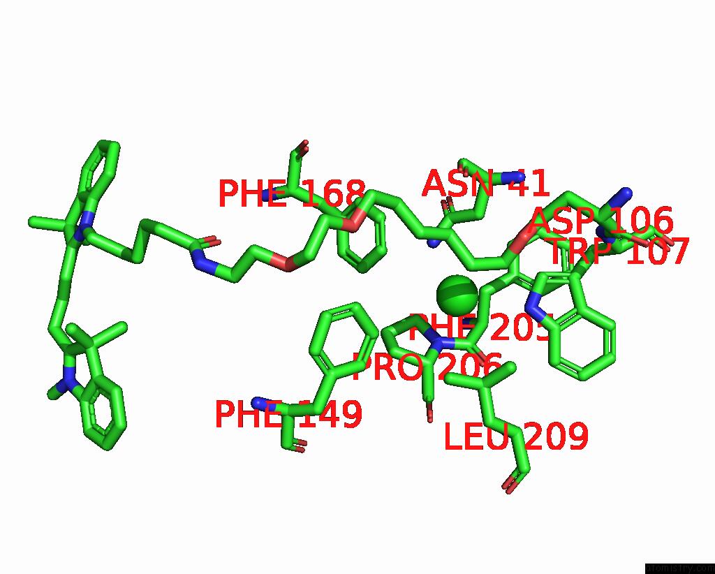
Mono view
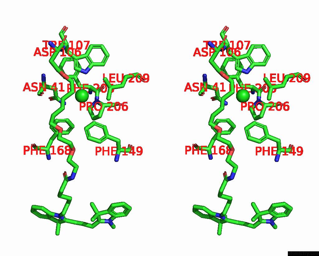
Stereo pair view
Mono view
Stereo pair view
A full contact list of Chlorine with other atoms in the Cl binding
site number 1 of X-Ray Structure of the Haloalkane Dehalogenase HALOTAG7 Labeled with A Chloroalkane CYANINE3 Fluorophore Substrate within 5.0Å range:
|
Reference:
L.Hellweg,
A.Edenhofer,
L.Barck,
M.C.Huppertz,
M.S.Frei,
M.Tarnawski,
A.Bergner,
B.Koch,
K.Johnsson,
J.Hiblot.
A General Method For the Development of Multicolor Biosensors with Large Dynamic Ranges. Nat.Chem.Biol. 2023.
ISSN: ESSN 1552-4469
PubMed: 37291200
DOI: 10.1038/S41589-023-01350-1
Page generated: Sun Jul 13 09:26:17 2025
ISSN: ESSN 1552-4469
PubMed: 37291200
DOI: 10.1038/S41589-023-01350-1
Last articles
Fe in 2YXOFe in 2YRS
Fe in 2YXC
Fe in 2YNM
Fe in 2YVJ
Fe in 2YP1
Fe in 2YU2
Fe in 2YU1
Fe in 2YQB
Fe in 2YOO