Chlorine »
PDB 162l-1ag9 »
1a7z »
Chlorine in PDB 1a7z: Crystal Structure of Actinomycin Z3
Protein crystallography data
The structure of Crystal Structure of Actinomycin Z3, PDB code: 1a7z
was solved by
M.Schafer,
with X-Ray Crystallography technique. A brief refinement statistics is given in the table below:
| Resolution Low / High (Å) | 32.45 / 0.95 |
| Space group | P 21 21 21 |
| Cell size a, b, c (Å), α, β, γ (°) | 14.803, 24.780, 65.059, 90.00, 90.00, 90.00 |
| R / Rfree (%) | 8 / 9.7 |
Chlorine Binding Sites:
The binding sites of Chlorine atom in the Crystal Structure of Actinomycin Z3
(pdb code 1a7z). This binding sites where shown within
5.0 Angstroms radius around Chlorine atom.
In total 3 binding sites of Chlorine where determined in the Crystal Structure of Actinomycin Z3, PDB code: 1a7z:
Jump to Chlorine binding site number: 1; 2; 3;
In total 3 binding sites of Chlorine where determined in the Crystal Structure of Actinomycin Z3, PDB code: 1a7z:
Jump to Chlorine binding site number: 1; 2; 3;
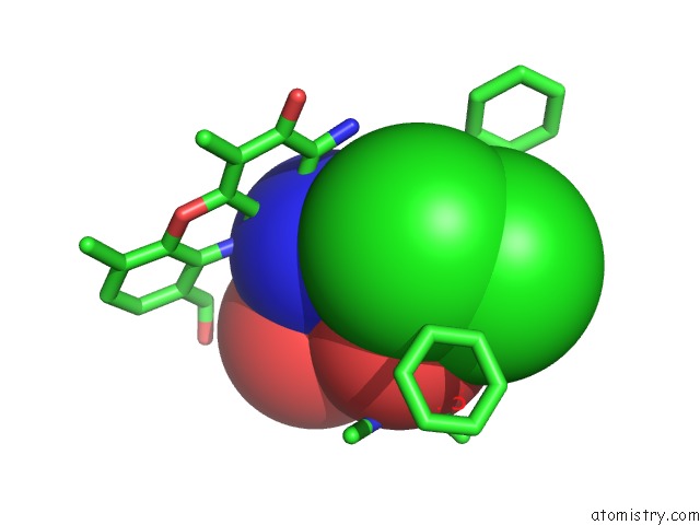
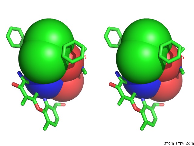
Chlorine binding site 1 out of 3 in 1a7z
Go back to
Chlorine binding site 1 out
of 3 in the Crystal Structure of Actinomycin Z3
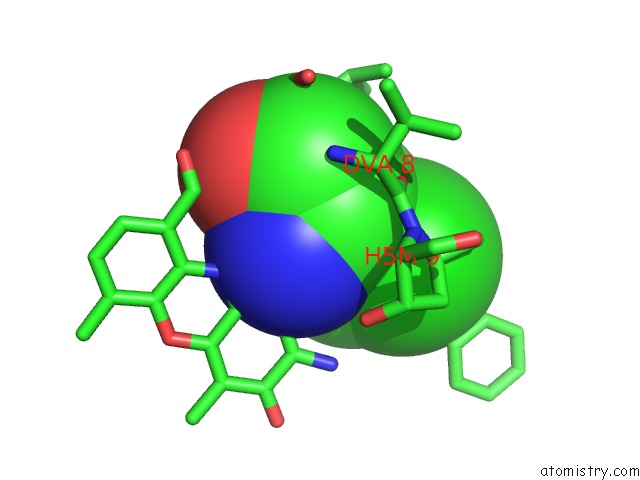
Mono view
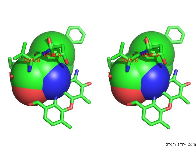
Stereo pair view
Mono view
Stereo pair view
A full contact list of Chlorine with other atoms in the Cl binding
site number 1 of Crystal Structure of Actinomycin Z3 within 5.0Å range:
|
Chlorine binding site 2 out of 3 in 1a7z
Go back to
Chlorine binding site 2 out
of 3 in the Crystal Structure of Actinomycin Z3
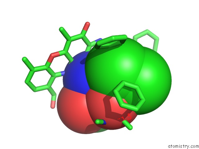
Mono view
Stereo pair view
Mono view
Stereo pair view
A full contact list of Chlorine with other atoms in the Cl binding
site number 2 of Crystal Structure of Actinomycin Z3 within 5.0Å range:
|
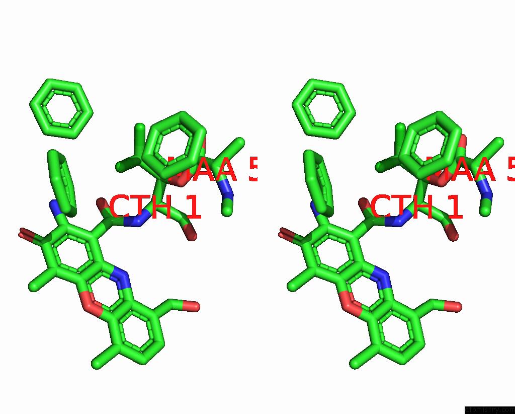
Chlorine binding site 3 out of 3 in 1a7z
Go back to
Chlorine binding site 3 out
of 3 in the Crystal Structure of Actinomycin Z3
Mono view
Stereo pair view
Mono view
Stereo pair view
A full contact list of Chlorine with other atoms in the Cl binding
site number 3 of Crystal Structure of Actinomycin Z3 within 5.0Å range:
|
Reference:
M.Schafer,
G.M.Sheldrick,
I.Bahner,
H.Lackner.
Crystal Structures of Actinomycin D and Actinomycin Z3. Angew.Chem.Int.Ed.Engl. V. 37 2381 1998.
ISSN: ESSN 1521-3773
PubMed: 29710967
DOI: 10.1002/(SICI)1521-3773(19980918)37:17<2381::AID-ANIE2381>3.0.CO;2-L
Page generated: Fri Jul 19 20:56:11 2024
ISSN: ESSN 1521-3773
PubMed: 29710967
DOI: 10.1002/(SICI)1521-3773(19980918)37:17<2381::AID-ANIE2381>3.0.CO;2-L
Last articles
Zn in 9J0NZn in 9J0O
Zn in 9J0P
Zn in 9FJX
Zn in 9EKB
Zn in 9C0F
Zn in 9CAH
Zn in 9CH0
Zn in 9CH3
Zn in 9CH1