Chlorine »
PDB 1ahz-1bxz »
1b2y »
Chlorine in PDB 1b2y: Structure of Human Pancreatic Alpha-Amylase in Complex with the Carbohydrate Inhibitor Acarbose
Enzymatic activity of Structure of Human Pancreatic Alpha-Amylase in Complex with the Carbohydrate Inhibitor Acarbose
All present enzymatic activity of Structure of Human Pancreatic Alpha-Amylase in Complex with the Carbohydrate Inhibitor Acarbose:
3.2.1.1;
3.2.1.1;
Protein crystallography data
The structure of Structure of Human Pancreatic Alpha-Amylase in Complex with the Carbohydrate Inhibitor Acarbose, PDB code: 1b2y
was solved by
V.Nahoum,
F.Payan,
with X-Ray Crystallography technique. A brief refinement statistics is given in the table below:
| Resolution Low / High (Å) | 11.00 / 3.20 |
| Space group | P 21 21 21 |
| Cell size a, b, c (Å), α, β, γ (°) | 53.110, 75.100, 137.130, 90.00, 90.00, 90.00 |
| R / Rfree (%) | 19.1 / 21.7 |
Other elements in 1b2y:
The structure of Structure of Human Pancreatic Alpha-Amylase in Complex with the Carbohydrate Inhibitor Acarbose also contains other interesting chemical elements:
| Calcium | (Ca) | 1 atom |
Chlorine Binding Sites:
The binding sites of Chlorine atom in the Structure of Human Pancreatic Alpha-Amylase in Complex with the Carbohydrate Inhibitor Acarbose
(pdb code 1b2y). This binding sites where shown within
5.0 Angstroms radius around Chlorine atom.
In total only one binding site of Chlorine was determined in the Structure of Human Pancreatic Alpha-Amylase in Complex with the Carbohydrate Inhibitor Acarbose, PDB code: 1b2y:
In total only one binding site of Chlorine was determined in the Structure of Human Pancreatic Alpha-Amylase in Complex with the Carbohydrate Inhibitor Acarbose, PDB code: 1b2y:
Chlorine binding site 1 out of 1 in 1b2y
Go back to
Chlorine binding site 1 out
of 1 in the Structure of Human Pancreatic Alpha-Amylase in Complex with the Carbohydrate Inhibitor Acarbose
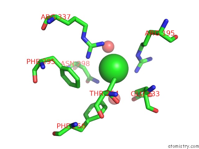
Mono view
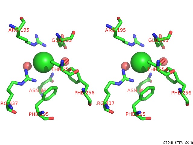
Stereo pair view
Mono view
Stereo pair view
A full contact list of Chlorine with other atoms in the Cl binding
site number 1 of Structure of Human Pancreatic Alpha-Amylase in Complex with the Carbohydrate Inhibitor Acarbose within 5.0Å range:
|
Reference:
V.Nahoum,
G.Roux,
V.Anton,
P.Rouge,
A.Puigserver,
H.Bischoff,
B.Henrissat,
F.Payan.
Crystal Structures of Human Pancreatic Alpha-Amylase in Complex with Carbohydrate and Proteinaceous Inhibitors. Biochem.J. V.Pt 1 201 2000.
ISSN: ISSN 0264-6021
PubMed: 10657258
DOI: 10.1042/0264-6021:3460201
Page generated: Fri Jul 19 21:02:18 2024
ISSN: ISSN 0264-6021
PubMed: 10657258
DOI: 10.1042/0264-6021:3460201
Last articles
Zn in 9MJ5Zn in 9HNW
Zn in 9G0L
Zn in 9FNE
Zn in 9DZN
Zn in 9E0I
Zn in 9D32
Zn in 9DAK
Zn in 8ZXC
Zn in 8ZUF