Chlorine »
PDB 1byz-1c6n »
1c3q »
Chlorine in PDB 1c3q: Crystal Structure of Native Thiazole Kinase in the Monoclinic Form
Enzymatic activity of Crystal Structure of Native Thiazole Kinase in the Monoclinic Form
All present enzymatic activity of Crystal Structure of Native Thiazole Kinase in the Monoclinic Form:
2.7.1.50;
2.7.1.50;
Protein crystallography data
The structure of Crystal Structure of Native Thiazole Kinase in the Monoclinic Form, PDB code: 1c3q
was solved by
N.Campobasso,
I.I.Mathews,
T.P.Begley,
S.E.Ealick,
with X-Ray Crystallography technique. A brief refinement statistics is given in the table below:
| Resolution Low / High (Å) | 20.00 / 2.00 |
| Space group | P 1 21 1 |
| Cell size a, b, c (Å), α, β, γ (°) | 53.920, 101.180, 73.330, 90.00, 95.26, 90.00 |
| R / Rfree (%) | 20.6 / 24.2 |
Chlorine Binding Sites:
The binding sites of Chlorine atom in the Crystal Structure of Native Thiazole Kinase in the Monoclinic Form
(pdb code 1c3q). This binding sites where shown within
5.0 Angstroms radius around Chlorine atom.
In total only one binding site of Chlorine was determined in the Crystal Structure of Native Thiazole Kinase in the Monoclinic Form, PDB code: 1c3q:
In total only one binding site of Chlorine was determined in the Crystal Structure of Native Thiazole Kinase in the Monoclinic Form, PDB code: 1c3q:
Chlorine binding site 1 out of 1 in 1c3q
Go back to
Chlorine binding site 1 out
of 1 in the Crystal Structure of Native Thiazole Kinase in the Monoclinic Form

Mono view
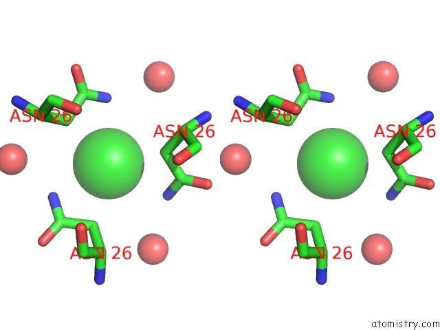
Stereo pair view
Mono view
Stereo pair view
A full contact list of Chlorine with other atoms in the Cl binding
site number 1 of Crystal Structure of Native Thiazole Kinase in the Monoclinic Form within 5.0Å range:
|
Reference:
N.Campobasso,
I.I.Mathews,
T.P.Begley,
S.E.Ealick.
Crystal Structure of 4-Methyl-5-Beta-Hydroxyethylthiazole Kinase From Bacillus Subtilis at 1.5 A Resolution. Biochemistry V. 39 7868 2000.
ISSN: ISSN 0006-2960
PubMed: 10891066
DOI: 10.1021/BI0000061
Page generated: Fri Jul 19 21:10:55 2024
ISSN: ISSN 0006-2960
PubMed: 10891066
DOI: 10.1021/BI0000061
Last articles
Zn in 9J0NZn in 9J0O
Zn in 9J0P
Zn in 9FJX
Zn in 9EKB
Zn in 9C0F
Zn in 9CAH
Zn in 9CH0
Zn in 9CH3
Zn in 9CH1