Chlorine »
PDB 1c6p-1cu5 »
1cb6 »
Chlorine in PDB 1cb6: Structure of Human Apolactoferrin at 2.0 A Resolution.
Protein crystallography data
The structure of Structure of Human Apolactoferrin at 2.0 A Resolution., PDB code: 1cb6
was solved by
G.B.Jameson,
B.F.Anderson,
G.E.Norris,
D.H.Thomas,
E.N.Baker,
with X-Ray Crystallography technique. A brief refinement statistics is given in the table below:
| Resolution Low / High (Å) | 10.00 / 2.00 |
| Space group | P 21 21 21 |
| Cell size a, b, c (Å), α, β, γ (°) | 152.090, 94.580, 55.790, 90.00, 90.00, 90.00 |
| R / Rfree (%) | 20.1 / 28.6 |
Chlorine Binding Sites:
The binding sites of Chlorine atom in the Structure of Human Apolactoferrin at 2.0 A Resolution.
(pdb code 1cb6). This binding sites where shown within
5.0 Angstroms radius around Chlorine atom.
In total 2 binding sites of Chlorine where determined in the Structure of Human Apolactoferrin at 2.0 A Resolution., PDB code: 1cb6:
Jump to Chlorine binding site number: 1; 2;
In total 2 binding sites of Chlorine where determined in the Structure of Human Apolactoferrin at 2.0 A Resolution., PDB code: 1cb6:
Jump to Chlorine binding site number: 1; 2;
Chlorine binding site 1 out of 2 in 1cb6
Go back to
Chlorine binding site 1 out
of 2 in the Structure of Human Apolactoferrin at 2.0 A Resolution.
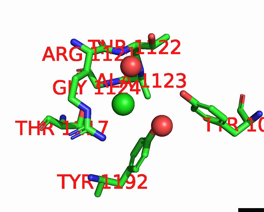
Mono view
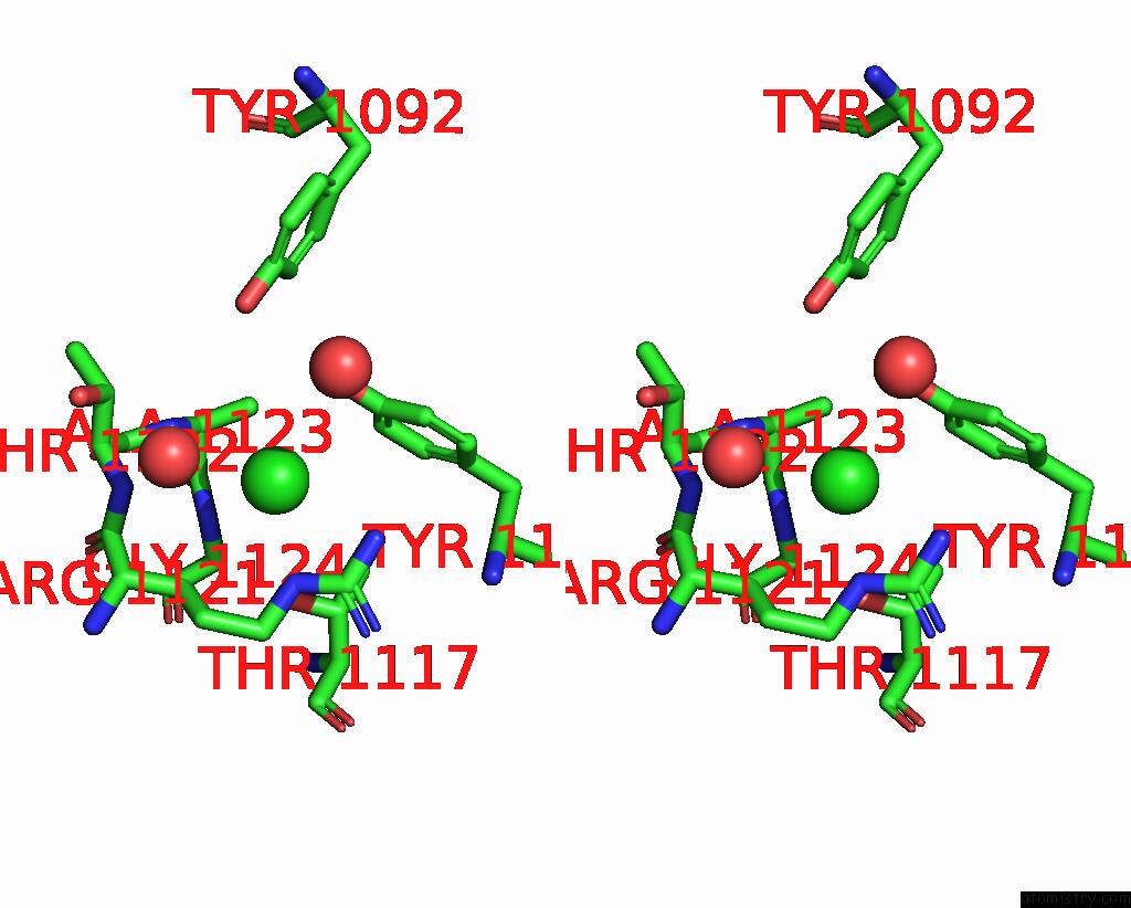
Stereo pair view
Mono view
Stereo pair view
A full contact list of Chlorine with other atoms in the Cl binding
site number 1 of Structure of Human Apolactoferrin at 2.0 A Resolution. within 5.0Å range:
|
Chlorine binding site 2 out of 2 in 1cb6
Go back to
Chlorine binding site 2 out
of 2 in the Structure of Human Apolactoferrin at 2.0 A Resolution.
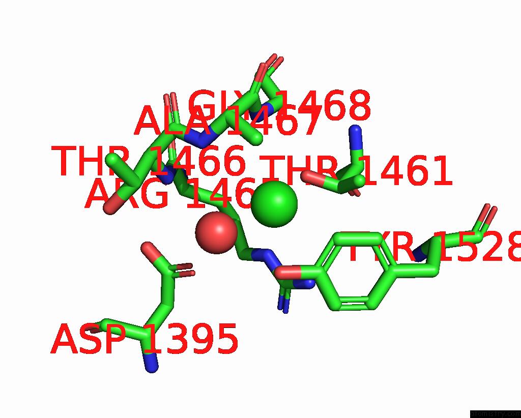
Mono view
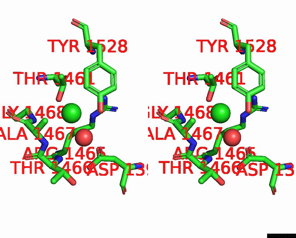
Stereo pair view
Mono view
Stereo pair view
A full contact list of Chlorine with other atoms in the Cl binding
site number 2 of Structure of Human Apolactoferrin at 2.0 A Resolution. within 5.0Å range:
|
Reference:
G.B.Jameson,
B.F.Anderson,
G.E.Norris,
D.H.Thomas,
E.N.Baker.
Structure of Human Apolactoferrin at 2.0 A Resolution. Refinement and Analysis of Ligand-Induced Conformational Change. Acta Crystallogr.,Sect.D V. 54 1319 1998.
ISSN: ISSN 0907-4449
PubMed: 10089508
DOI: 10.1107/S0907444998004417
Page generated: Thu Jul 10 16:30:31 2025
ISSN: ISSN 0907-4449
PubMed: 10089508
DOI: 10.1107/S0907444998004417
Last articles
Cl in 7YYGCl in 7YWK
Cl in 7YXV
Cl in 7YXM
Cl in 7YUZ
Cl in 7YVV
Cl in 7YWJ
Cl in 7YWB
Cl in 7YRK
Cl in 7YUG