Chlorine »
PDB 1c6p-1cu5 »
1ch0 »
Chlorine in PDB 1ch0: Rnase T1 Variant with Altered Guanine Binding Segment
Enzymatic activity of Rnase T1 Variant with Altered Guanine Binding Segment
All present enzymatic activity of Rnase T1 Variant with Altered Guanine Binding Segment:
3.1.27.3;
3.1.27.3;
Protein crystallography data
The structure of Rnase T1 Variant with Altered Guanine Binding Segment, PDB code: 1ch0
was solved by
K.Hoeschler,
H.Hoier,
P.Orth,
B.Hubner,
W.Saenger,
U.Hahn,
with X-Ray Crystallography technique. A brief refinement statistics is given in the table below:
| Resolution Low / High (Å) | 35.60 / 2.30 |
| Space group | P 21 21 21 |
| Cell size a, b, c (Å), α, β, γ (°) | 39.820, 48.850, 158.770, 90.00, 90.00, 90.00 |
| R / Rfree (%) | 19.8 / 24.1 |
Other elements in 1ch0:
The structure of Rnase T1 Variant with Altered Guanine Binding Segment also contains other interesting chemical elements:
| Calcium | (Ca) | 4 atoms |
Chlorine Binding Sites:
The binding sites of Chlorine atom in the Rnase T1 Variant with Altered Guanine Binding Segment
(pdb code 1ch0). This binding sites where shown within
5.0 Angstroms radius around Chlorine atom.
In total only one binding site of Chlorine was determined in the Rnase T1 Variant with Altered Guanine Binding Segment, PDB code: 1ch0:
In total only one binding site of Chlorine was determined in the Rnase T1 Variant with Altered Guanine Binding Segment, PDB code: 1ch0:
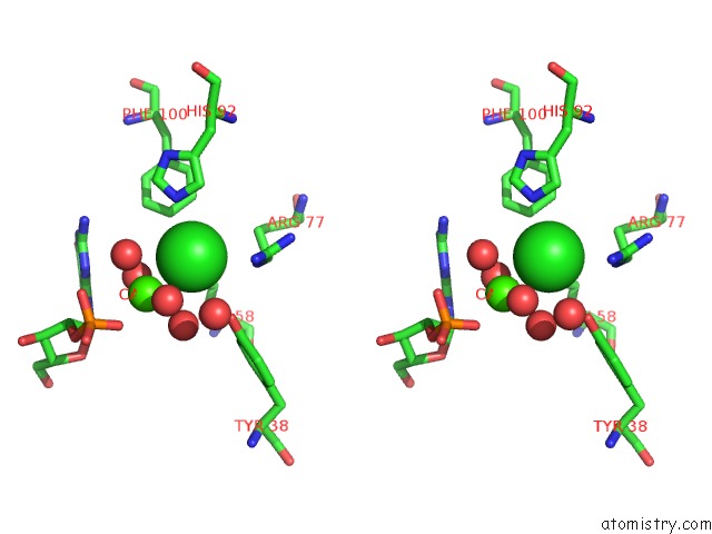
Chlorine binding site 1 out of 1 in 1ch0
Go back to
Chlorine binding site 1 out
of 1 in the Rnase T1 Variant with Altered Guanine Binding Segment

Mono view
Stereo pair view
Mono view
Stereo pair view
A full contact list of Chlorine with other atoms in the Cl binding
site number 1 of Rnase T1 Variant with Altered Guanine Binding Segment within 5.0Å range:
|
Reference:
K.Hoschler,
H.Hoier,
B.Hubner,
W.Saenger,
P.Orth,
U.Hahn.
Structural Analysis of An Rnase T1 Variant with An Altered Guanine Binding Segment. J.Mol.Biol. V. 294 1231 1999.
ISSN: ISSN 0022-2836
PubMed: 10600381
DOI: 10.1006/JMBI.1999.3324
Page generated: Thu Jul 10 16:30:56 2025
ISSN: ISSN 0022-2836
PubMed: 10600381
DOI: 10.1006/JMBI.1999.3324
Last articles
Fe in 2YXOFe in 2YRS
Fe in 2YXC
Fe in 2YNM
Fe in 2YVJ
Fe in 2YP1
Fe in 2YU2
Fe in 2YU1
Fe in 2YQB
Fe in 2YOO