Chlorine »
PDB 1cu6-1dhi »
1cx7 »
Chlorine in PDB 1cx7: T4 Lysozyme Methionine Core Mutant
Enzymatic activity of T4 Lysozyme Methionine Core Mutant
All present enzymatic activity of T4 Lysozyme Methionine Core Mutant:
3.2.1.17;
3.2.1.17;
Protein crystallography data
The structure of T4 Lysozyme Methionine Core Mutant, PDB code: 1cx7
was solved by
N.C.Gassner,
W.A.Baase,
J.Lindstrom,
J.Lu,
B.W.Matthews,
with X-Ray Crystallography technique. A brief refinement statistics is given in the table below:
| Resolution Low / High (Å) | 30.00 / 1.94 |
| Space group | P 32 2 1 |
| Cell size a, b, c (Å), α, β, γ (°) | 61.310, 61.310, 96.490, 90.00, 90.00, 120.00 |
| R / Rfree (%) | n/a / n/a |
Chlorine Binding Sites:
The binding sites of Chlorine atom in the T4 Lysozyme Methionine Core Mutant
(pdb code 1cx7). This binding sites where shown within
5.0 Angstroms radius around Chlorine atom.
In total 2 binding sites of Chlorine where determined in the T4 Lysozyme Methionine Core Mutant, PDB code: 1cx7:
Jump to Chlorine binding site number: 1; 2;
In total 2 binding sites of Chlorine where determined in the T4 Lysozyme Methionine Core Mutant, PDB code: 1cx7:
Jump to Chlorine binding site number: 1; 2;
Chlorine binding site 1 out of 2 in 1cx7
Go back to
Chlorine binding site 1 out
of 2 in the T4 Lysozyme Methionine Core Mutant
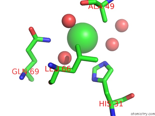
Mono view
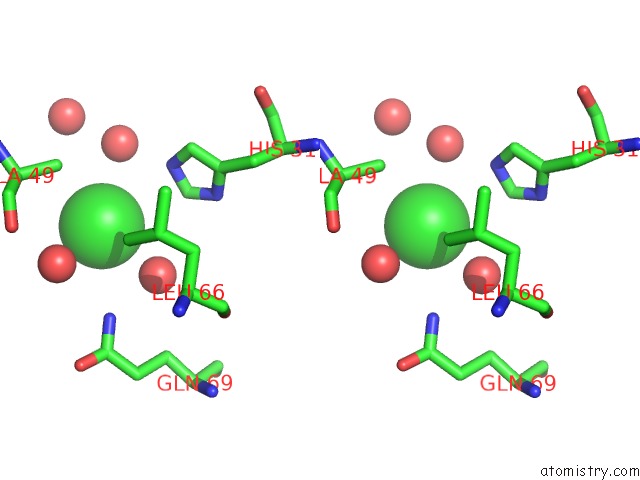
Stereo pair view
Mono view
Stereo pair view
A full contact list of Chlorine with other atoms in the Cl binding
site number 1 of T4 Lysozyme Methionine Core Mutant within 5.0Å range:
|
Chlorine binding site 2 out of 2 in 1cx7
Go back to
Chlorine binding site 2 out
of 2 in the T4 Lysozyme Methionine Core Mutant
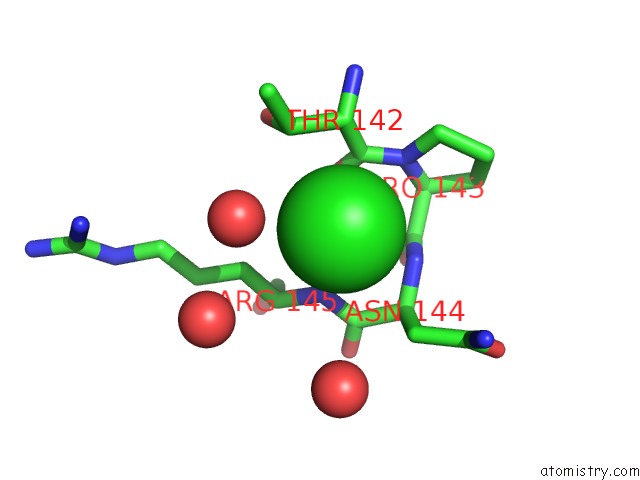
Mono view
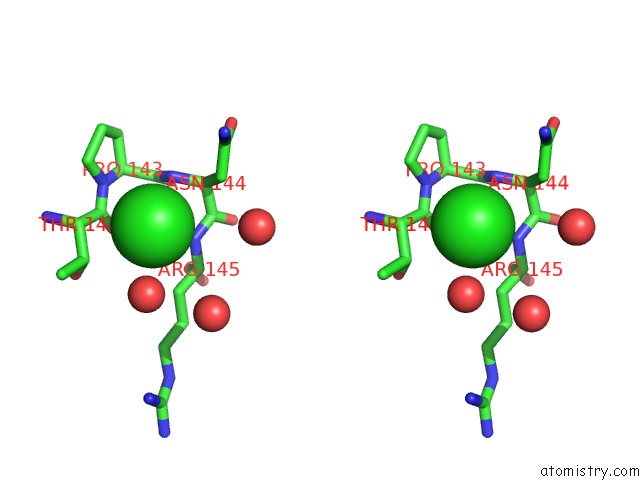
Stereo pair view
Mono view
Stereo pair view
A full contact list of Chlorine with other atoms in the Cl binding
site number 2 of T4 Lysozyme Methionine Core Mutant within 5.0Å range:
|
Reference:
N.C.Gassner,
B.W.Matthews.
Use of Differentially Substituted Selenomethionine Proteins in X-Ray Structure Determination. Acta Crystallogr.,Sect.D V. 55 1967 1999.
ISSN: ISSN 0907-4449
PubMed: 10666571
DOI: 10.1021/BI9915519
Page generated: Fri Jul 19 21:30:19 2024
ISSN: ISSN 0907-4449
PubMed: 10666571
DOI: 10.1021/BI9915519
Last articles
Zn in 9J0NZn in 9J0O
Zn in 9J0P
Zn in 9FJX
Zn in 9EKB
Zn in 9C0F
Zn in 9CAH
Zn in 9CH0
Zn in 9CH3
Zn in 9CH1