Chlorine »
PDB 1dhj-1e2y »
1diq »
Chlorine in PDB 1diq: Crystal Structure of P-Cresol Methylhydroxylase with Substrate Bound
Enzymatic activity of Crystal Structure of P-Cresol Methylhydroxylase with Substrate Bound
All present enzymatic activity of Crystal Structure of P-Cresol Methylhydroxylase with Substrate Bound:
1.17.99.1;
1.17.99.1;
Protein crystallography data
The structure of Crystal Structure of P-Cresol Methylhydroxylase with Substrate Bound, PDB code: 1diq
was solved by
L.M.Cunane,
Z.W.Chen,
N.Shamala,
F.S.Mathews,
C.S.Cronin,
W.S.Mcintire,
with X-Ray Crystallography technique. A brief refinement statistics is given in the table below:
| Resolution Low / High (Å) | 25.00 / 2.75 |
| Space group | P 21 21 21 |
| Cell size a, b, c (Å), α, β, γ (°) | 139.100, 130.500, 74.400, 90.00, 90.00, 90.00 |
| R / Rfree (%) | 13.3 / 18.3 |
Other elements in 1diq:
The structure of Crystal Structure of P-Cresol Methylhydroxylase with Substrate Bound also contains other interesting chemical elements:
| Iron | (Fe) | 2 atoms |
Chlorine Binding Sites:
The binding sites of Chlorine atom in the Crystal Structure of P-Cresol Methylhydroxylase with Substrate Bound
(pdb code 1diq). This binding sites where shown within
5.0 Angstroms radius around Chlorine atom.
In total 2 binding sites of Chlorine where determined in the Crystal Structure of P-Cresol Methylhydroxylase with Substrate Bound, PDB code: 1diq:
Jump to Chlorine binding site number: 1; 2;
In total 2 binding sites of Chlorine where determined in the Crystal Structure of P-Cresol Methylhydroxylase with Substrate Bound, PDB code: 1diq:
Jump to Chlorine binding site number: 1; 2;
Chlorine binding site 1 out of 2 in 1diq
Go back to
Chlorine binding site 1 out
of 2 in the Crystal Structure of P-Cresol Methylhydroxylase with Substrate Bound
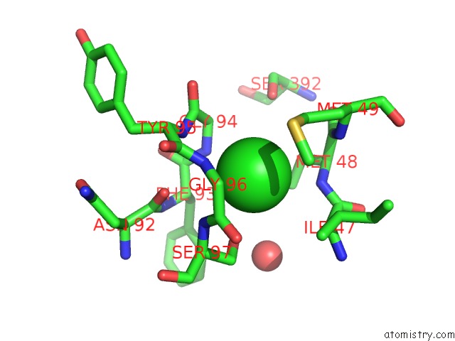
Mono view
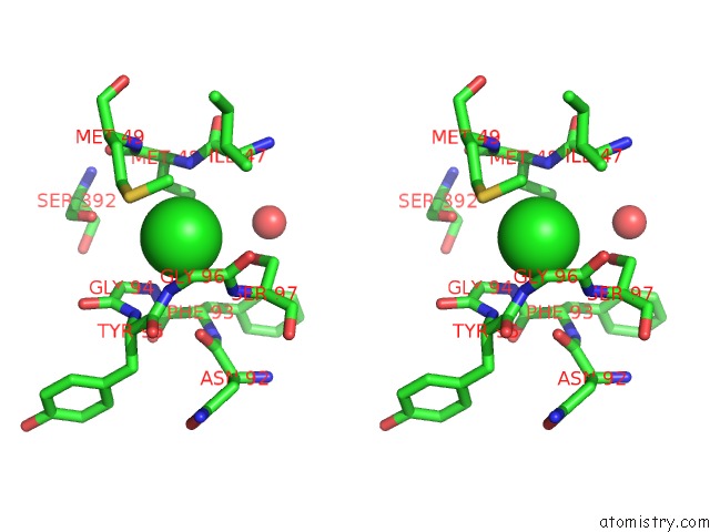
Stereo pair view
Mono view
Stereo pair view
A full contact list of Chlorine with other atoms in the Cl binding
site number 1 of Crystal Structure of P-Cresol Methylhydroxylase with Substrate Bound within 5.0Å range:
|
Chlorine binding site 2 out of 2 in 1diq
Go back to
Chlorine binding site 2 out
of 2 in the Crystal Structure of P-Cresol Methylhydroxylase with Substrate Bound
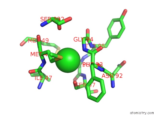
Mono view
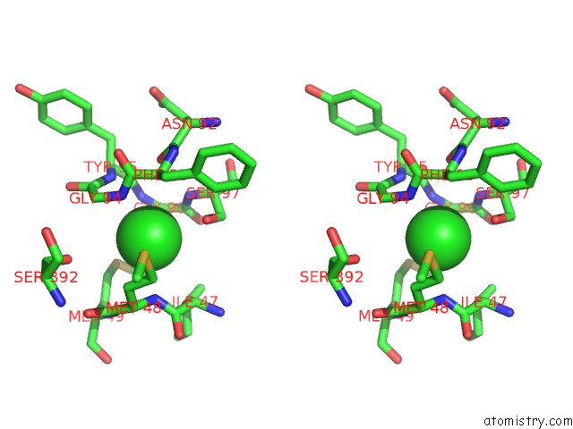
Stereo pair view
Mono view
Stereo pair view
A full contact list of Chlorine with other atoms in the Cl binding
site number 2 of Crystal Structure of P-Cresol Methylhydroxylase with Substrate Bound within 5.0Å range:
|
Reference:
L.M.Cunane,
Z.W.Chen,
N.Shamala,
F.S.Mathews,
C.N.Cronin,
W.S.Mcintire.
Structures of the Flavocytochrome P-Cresol Methylhydroxylase and Its Enzyme-Substrate Complex: Gated Substrate Entry and Proton Relays Support the Proposed Catalytic Mechanism. J.Mol.Biol. V. 295 357 2000.
ISSN: ISSN 0022-2836
PubMed: 10623531
DOI: 10.1006/JMBI.1999.3290
Page generated: Fri Jul 19 21:35:05 2024
ISSN: ISSN 0022-2836
PubMed: 10623531
DOI: 10.1006/JMBI.1999.3290
Last articles
Zn in 9J0NZn in 9J0O
Zn in 9J0P
Zn in 9FJX
Zn in 9EKB
Zn in 9C0F
Zn in 9CAH
Zn in 9CH0
Zn in 9CH3
Zn in 9CH1