Chlorine »
PDB 1go6-1hav »
1hav »
Chlorine in PDB 1hav: Hepatitis A Virus 3C Proteinase
Enzymatic activity of Hepatitis A Virus 3C Proteinase
All present enzymatic activity of Hepatitis A Virus 3C Proteinase:
2.7.7.48;
2.7.7.48;
Protein crystallography data
The structure of Hepatitis A Virus 3C Proteinase, PDB code: 1hav
was solved by
E.M.Bergmann,
M.N.G.James,
with X-Ray Crystallography technique. A brief refinement statistics is given in the table below:
| Resolution Low / High (Å) | 20.00 / 2.00 |
| Space group | P 1 |
| Cell size a, b, c (Å), α, β, γ (°) | 53.600, 53.550, 53.200, 99.08, 129.00, 103.31 |
| R / Rfree (%) | 21.1 / 26.5 |
Chlorine Binding Sites:
The binding sites of Chlorine atom in the Hepatitis A Virus 3C Proteinase
(pdb code 1hav). This binding sites where shown within
5.0 Angstroms radius around Chlorine atom.
In total only one binding site of Chlorine was determined in the Hepatitis A Virus 3C Proteinase, PDB code: 1hav:
In total only one binding site of Chlorine was determined in the Hepatitis A Virus 3C Proteinase, PDB code: 1hav:
Chlorine binding site 1 out of 1 in 1hav
Go back to
Chlorine binding site 1 out
of 1 in the Hepatitis A Virus 3C Proteinase
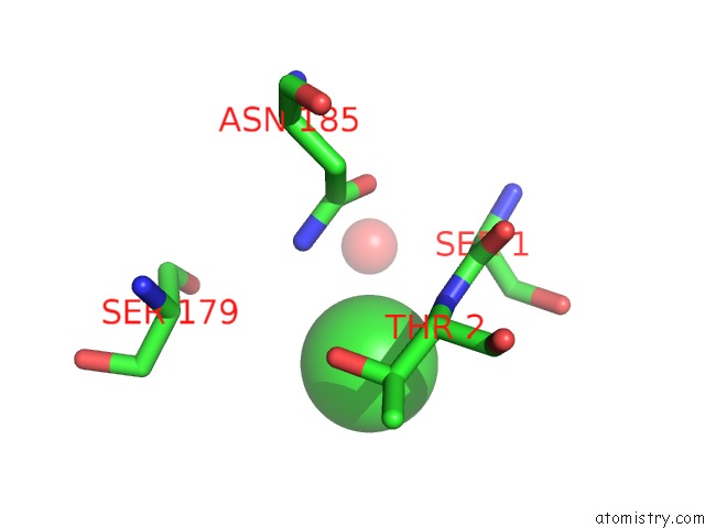
Mono view
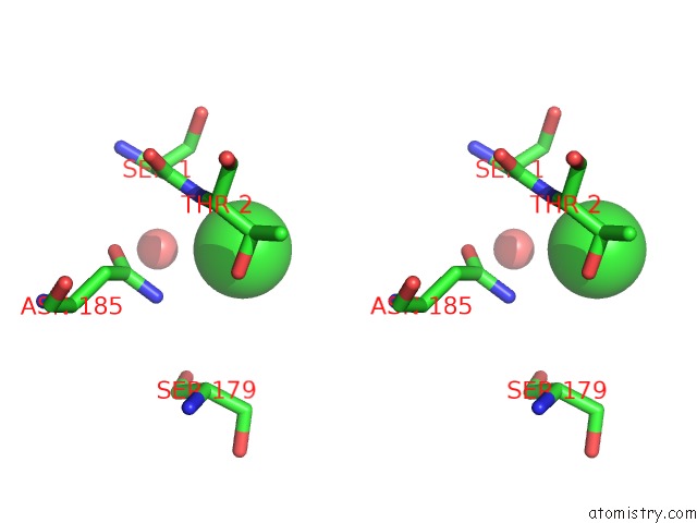
Stereo pair view
Mono view
Stereo pair view
A full contact list of Chlorine with other atoms in the Cl binding
site number 1 of Hepatitis A Virus 3C Proteinase within 5.0Å range:
|
Reference:
E.M.Bergmann,
S.C.Mosimann,
M.M.Chernaia,
B.A.Malcolm,
M.N.James.
The Refined Crystal Structure of the 3C Gene Product From Hepatitis A Virus: Specific Proteinase Activity and Rna Recognition. J.Virol. V. 71 2436 1997.
ISSN: ISSN 0022-538X
PubMed: 9032381
Page generated: Fri Jul 19 22:26:52 2024
ISSN: ISSN 0022-538X
PubMed: 9032381
Last articles
Zn in 9J0NZn in 9J0O
Zn in 9J0P
Zn in 9FJX
Zn in 9EKB
Zn in 9C0F
Zn in 9CAH
Zn in 9CH0
Zn in 9CH3
Zn in 9CH1