Chlorine »
PDB 1hba-1i3k »
1hnv »
Chlorine in PDB 1hnv: Structure of Hiv-1 Rt(Slash)Tibo R 86183 Complex Reveals Similarity in the Binding of Diverse Nonnucleoside Inhibitors
Enzymatic activity of Structure of Hiv-1 Rt(Slash)Tibo R 86183 Complex Reveals Similarity in the Binding of Diverse Nonnucleoside Inhibitors
All present enzymatic activity of Structure of Hiv-1 Rt(Slash)Tibo R 86183 Complex Reveals Similarity in the Binding of Diverse Nonnucleoside Inhibitors:
2.7.7.49;
2.7.7.49;
Protein crystallography data
The structure of Structure of Hiv-1 Rt(Slash)Tibo R 86183 Complex Reveals Similarity in the Binding of Diverse Nonnucleoside Inhibitors, PDB code: 1hnv
was solved by
K.Das,
J.Ding,
E.Arnold,
with X-Ray Crystallography technique. A brief refinement statistics is given in the table below:
| Resolution Low / High (Å) | 10.00 / 3.00 |
| Space group | C 1 2 1 |
| Cell size a, b, c (Å), α, β, γ (°) | 227.200, 70.200, 105.700, 90.00, 105.60, 90.00 |
| R / Rfree (%) | 24.9 / 35.6 |
Chlorine Binding Sites:
The binding sites of Chlorine atom in the Structure of Hiv-1 Rt(Slash)Tibo R 86183 Complex Reveals Similarity in the Binding of Diverse Nonnucleoside Inhibitors
(pdb code 1hnv). This binding sites where shown within
5.0 Angstroms radius around Chlorine atom.
In total only one binding site of Chlorine was determined in the Structure of Hiv-1 Rt(Slash)Tibo R 86183 Complex Reveals Similarity in the Binding of Diverse Nonnucleoside Inhibitors, PDB code: 1hnv:
In total only one binding site of Chlorine was determined in the Structure of Hiv-1 Rt(Slash)Tibo R 86183 Complex Reveals Similarity in the Binding of Diverse Nonnucleoside Inhibitors, PDB code: 1hnv:
Chlorine binding site 1 out of 1 in 1hnv
Go back to
Chlorine binding site 1 out
of 1 in the Structure of Hiv-1 Rt(Slash)Tibo R 86183 Complex Reveals Similarity in the Binding of Diverse Nonnucleoside Inhibitors
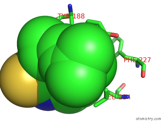
Mono view
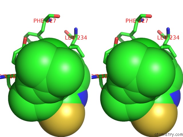
Stereo pair view
Mono view
Stereo pair view
A full contact list of Chlorine with other atoms in the Cl binding
site number 1 of Structure of Hiv-1 Rt(Slash)Tibo R 86183 Complex Reveals Similarity in the Binding of Diverse Nonnucleoside Inhibitors within 5.0Å range:
|
Reference:
J.Ding,
K.Das,
H.Moereels,
L.Koymans,
K.Andries,
P.A.Janssen,
S.H.Hughes,
E.Arnold.
Structure of Hiv-1 Rt/Tibo R 86183 Complex Reveals Similarity in the Binding of Diverse Nonnucleoside Inhibitors. Nat.Struct.Biol. V. 2 407 1995.
ISSN: ISSN 1072-8368
PubMed: 7545077
DOI: 10.1038/NSB0595-407
Page generated: Fri Jul 19 22:32:42 2024
ISSN: ISSN 1072-8368
PubMed: 7545077
DOI: 10.1038/NSB0595-407
Last articles
Zn in 9J0NZn in 9J0O
Zn in 9J0P
Zn in 9FJX
Zn in 9EKB
Zn in 9C0F
Zn in 9CAH
Zn in 9CH0
Zn in 9CH3
Zn in 9CH1