Chlorine »
PDB 1hba-1i3k »
1hw6 »
Chlorine in PDB 1hw6: Crystal Structure of Apo-2,5-Diketo-D-Gluconate Reductase
Protein crystallography data
The structure of Crystal Structure of Apo-2,5-Diketo-D-Gluconate Reductase, PDB code: 1hw6
was solved by
G.Sanli,
M.Blaber,
with X-Ray Crystallography technique. A brief refinement statistics is given in the table below:
| Resolution Low / High (Å) | 27.00 / 1.90 |
| Space group | P 21 21 21 |
| Cell size a, b, c (Å), α, β, γ (°) | 53.030, 53.940, 89.750, 90.00, 90.00, 90.00 |
| R / Rfree (%) | 20 / 26.8 |
Other elements in 1hw6:
The structure of Crystal Structure of Apo-2,5-Diketo-D-Gluconate Reductase also contains other interesting chemical elements:
| Magnesium | (Mg) | 2 atoms |
Chlorine Binding Sites:
The binding sites of Chlorine atom in the Crystal Structure of Apo-2,5-Diketo-D-Gluconate Reductase
(pdb code 1hw6). This binding sites where shown within
5.0 Angstroms radius around Chlorine atom.
In total 2 binding sites of Chlorine where determined in the Crystal Structure of Apo-2,5-Diketo-D-Gluconate Reductase, PDB code: 1hw6:
Jump to Chlorine binding site number: 1; 2;
In total 2 binding sites of Chlorine where determined in the Crystal Structure of Apo-2,5-Diketo-D-Gluconate Reductase, PDB code: 1hw6:
Jump to Chlorine binding site number: 1; 2;
Chlorine binding site 1 out of 2 in 1hw6
Go back to
Chlorine binding site 1 out
of 2 in the Crystal Structure of Apo-2,5-Diketo-D-Gluconate Reductase
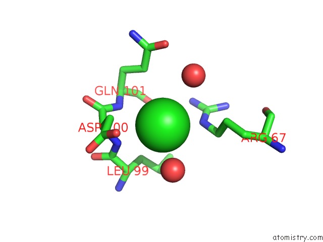
Mono view
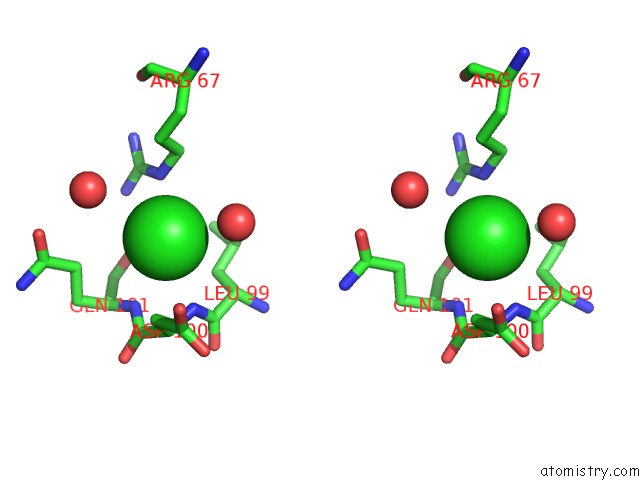
Stereo pair view
Mono view
Stereo pair view
A full contact list of Chlorine with other atoms in the Cl binding
site number 1 of Crystal Structure of Apo-2,5-Diketo-D-Gluconate Reductase within 5.0Å range:
|
Chlorine binding site 2 out of 2 in 1hw6
Go back to
Chlorine binding site 2 out
of 2 in the Crystal Structure of Apo-2,5-Diketo-D-Gluconate Reductase
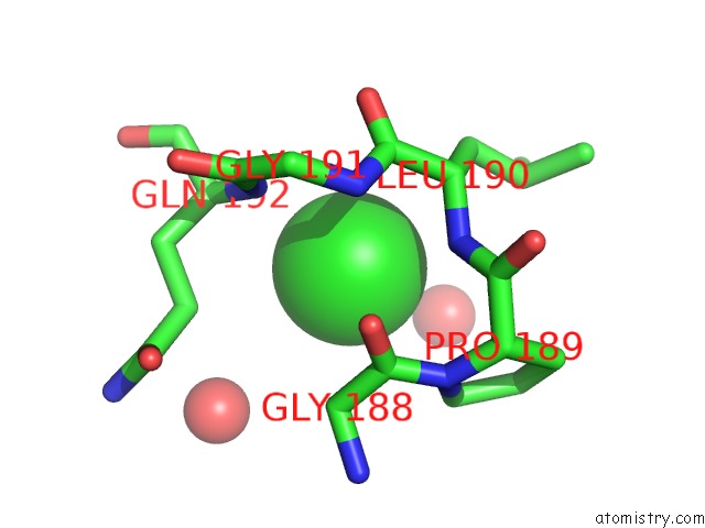
Mono view
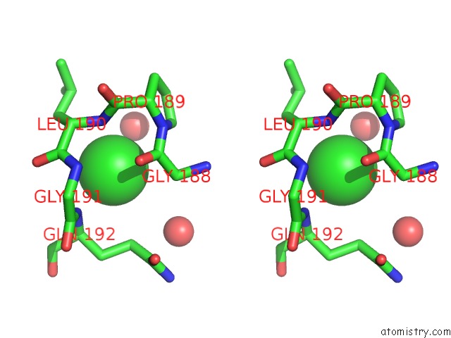
Stereo pair view
Mono view
Stereo pair view
A full contact list of Chlorine with other atoms in the Cl binding
site number 2 of Crystal Structure of Apo-2,5-Diketo-D-Gluconate Reductase within 5.0Å range:
|
Reference:
G.Sanli,
M.Blaber.
Structural Assembly of the Active Site in An Aldo-Keto Reductase By Nadph Cofactor. J.Mol.Biol. V. 309 1209 2001.
ISSN: ISSN 0022-2836
PubMed: 11399090
DOI: 10.1006/JMBI.2001.4739
Page generated: Fri Jul 19 22:35:20 2024
ISSN: ISSN 0022-2836
PubMed: 11399090
DOI: 10.1006/JMBI.2001.4739
Last articles
Zn in 9J0NZn in 9J0O
Zn in 9J0P
Zn in 9FJX
Zn in 9EKB
Zn in 9C0F
Zn in 9CAH
Zn in 9CH0
Zn in 9CH3
Zn in 9CH1