Chlorine »
PDB 1hba-1i3k »
1hyu »
Chlorine in PDB 1hyu: Crystal Structure of Intact Ahpf
Protein crystallography data
The structure of Crystal Structure of Intact Ahpf, PDB code: 1hyu
was solved by
Z.A.Wood,
L.B.Poole,
P.A.Karplus,
with X-Ray Crystallography technique. A brief refinement statistics is given in the table below:
| Resolution Low / High (Å) | 29.88 / 2.00 |
| Space group | P 21 21 2 |
| Cell size a, b, c (Å), α, β, γ (°) | 102.200, 139.160, 39.060, 90.00, 90.00, 90.00 |
| R / Rfree (%) | 18.2 / 23.5 |
Chlorine Binding Sites:
The binding sites of Chlorine atom in the Crystal Structure of Intact Ahpf
(pdb code 1hyu). This binding sites where shown within
5.0 Angstroms radius around Chlorine atom.
In total only one binding site of Chlorine was determined in the Crystal Structure of Intact Ahpf, PDB code: 1hyu:
In total only one binding site of Chlorine was determined in the Crystal Structure of Intact Ahpf, PDB code: 1hyu:
Chlorine binding site 1 out of 1 in 1hyu
Go back to
Chlorine binding site 1 out
of 1 in the Crystal Structure of Intact Ahpf
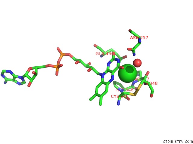
Mono view
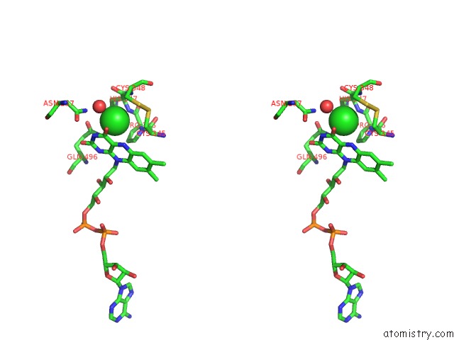
Stereo pair view
Mono view
Stereo pair view
A full contact list of Chlorine with other atoms in the Cl binding
site number 1 of Crystal Structure of Intact Ahpf within 5.0Å range:
|
Reference:
Z.A.Wood,
L.B.Poole,
P.A.Karplus.
Structure of Intact Ahpf Reveals A Mirrored Thioredoxin-Like Active Site and Implies Large Domain Rotations During Catalysis. Biochemistry V. 40 3900 2001.
ISSN: ISSN 0006-2960
PubMed: 11300769
DOI: 10.1021/BI002765P
Page generated: Fri Jul 19 22:36:50 2024
ISSN: ISSN 0006-2960
PubMed: 11300769
DOI: 10.1021/BI002765P
Last articles
Zn in 9J0NZn in 9J0O
Zn in 9J0P
Zn in 9FJX
Zn in 9EKB
Zn in 9C0F
Zn in 9CAH
Zn in 9CH0
Zn in 9CH3
Zn in 9CH1