Chlorine »
PDB 1i3l-1iqi »
1iep »
Chlorine in PDB 1iep: Crystal Structure of the C-Abl Kinase Domain in Complex with Sti-571.
Enzymatic activity of Crystal Structure of the C-Abl Kinase Domain in Complex with Sti-571.
All present enzymatic activity of Crystal Structure of the C-Abl Kinase Domain in Complex with Sti-571.:
2.7.1.112;
2.7.1.112;
Protein crystallography data
The structure of Crystal Structure of the C-Abl Kinase Domain in Complex with Sti-571., PDB code: 1iep
was solved by
B.Nagar,
W.Bornmann,
T.Schindler,
B.Clarkson,
J.Kuriyan,
with X-Ray Crystallography technique. A brief refinement statistics is given in the table below:
| Resolution Low / High (Å) | 29.14 / 2.10 |
| Space group | F 2 2 2 |
| Cell size a, b, c (Å), α, β, γ (°) | 112.885, 147.371, 153.442, 90.00, 90.00, 90.00 |
| R / Rfree (%) | 23.1 / 26.2 |
Chlorine Binding Sites:
The binding sites of Chlorine atom in the Crystal Structure of the C-Abl Kinase Domain in Complex with Sti-571.
(pdb code 1iep). This binding sites where shown within
5.0 Angstroms radius around Chlorine atom.
In total 6 binding sites of Chlorine where determined in the Crystal Structure of the C-Abl Kinase Domain in Complex with Sti-571., PDB code: 1iep:
Jump to Chlorine binding site number: 1; 2; 3; 4; 5; 6;
In total 6 binding sites of Chlorine where determined in the Crystal Structure of the C-Abl Kinase Domain in Complex with Sti-571., PDB code: 1iep:
Jump to Chlorine binding site number: 1; 2; 3; 4; 5; 6;
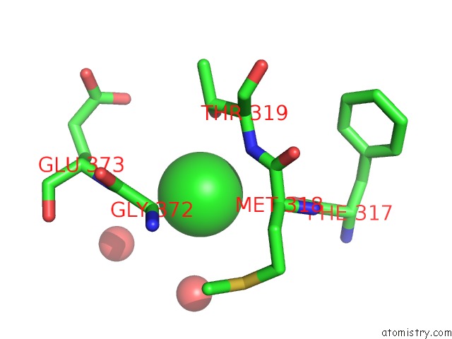
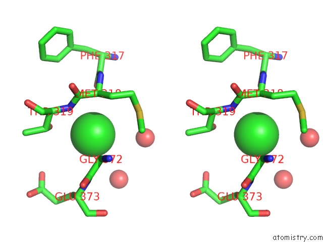
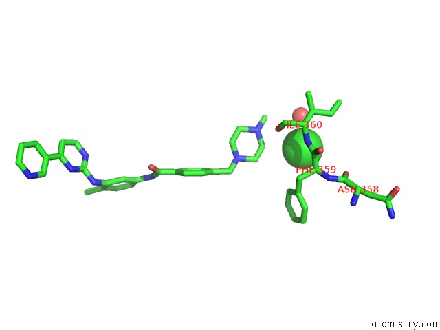
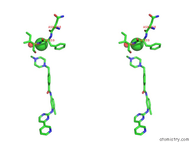
Chlorine binding site 1 out of 6 in 1iep
Go back to
Chlorine binding site 1 out
of 6 in the Crystal Structure of the C-Abl Kinase Domain in Complex with Sti-571.
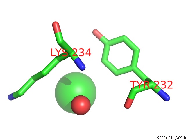
Mono view
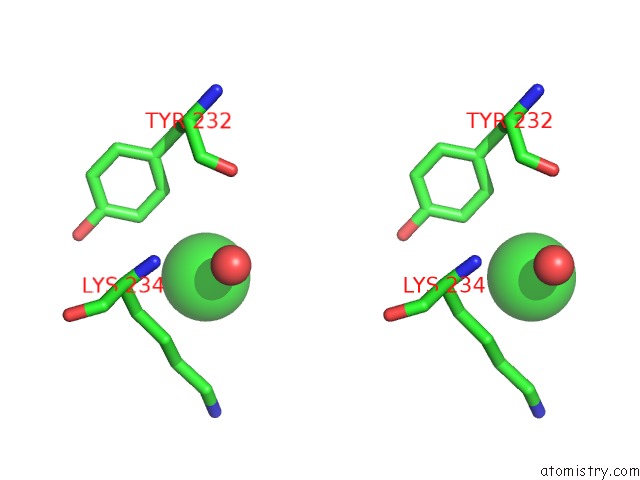
Stereo pair view
Mono view
Stereo pair view
A full contact list of Chlorine with other atoms in the Cl binding
site number 1 of Crystal Structure of the C-Abl Kinase Domain in Complex with Sti-571. within 5.0Å range:
|
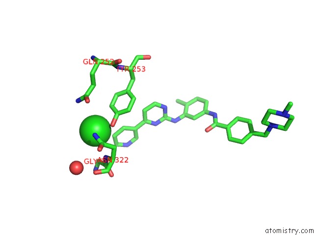
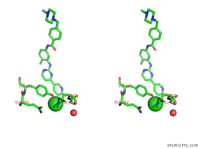
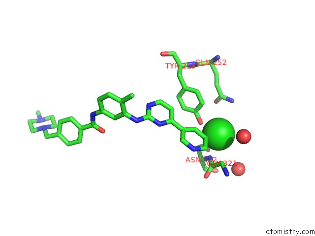
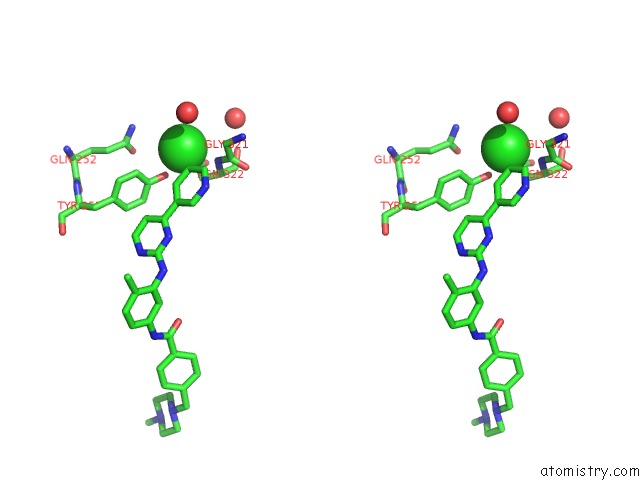
Chlorine binding site 2 out of 6 in 1iep
Go back to
Chlorine binding site 2 out
of 6 in the Crystal Structure of the C-Abl Kinase Domain in Complex with Sti-571.
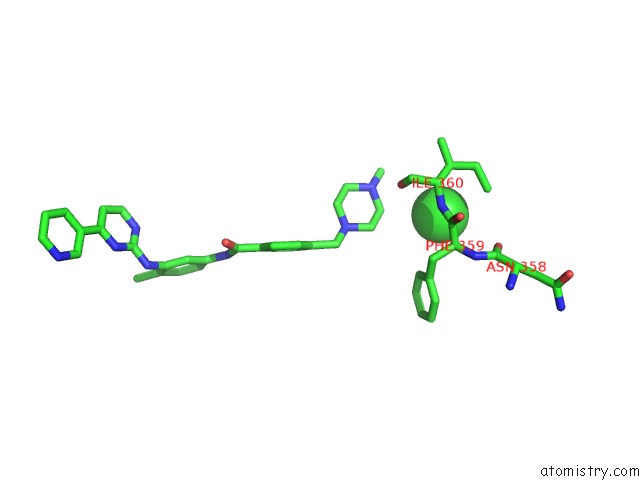
Mono view
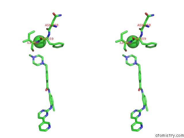
Stereo pair view
Mono view
Stereo pair view
A full contact list of Chlorine with other atoms in the Cl binding
site number 2 of Crystal Structure of the C-Abl Kinase Domain in Complex with Sti-571. within 5.0Å range:
|
Chlorine binding site 3 out of 6 in 1iep
Go back to
Chlorine binding site 3 out
of 6 in the Crystal Structure of the C-Abl Kinase Domain in Complex with Sti-571.
Mono view
Stereo pair view
Mono view
Stereo pair view
A full contact list of Chlorine with other atoms in the Cl binding
site number 3 of Crystal Structure of the C-Abl Kinase Domain in Complex with Sti-571. within 5.0Å range:
|
Chlorine binding site 4 out of 6 in 1iep
Go back to
Chlorine binding site 4 out
of 6 in the Crystal Structure of the C-Abl Kinase Domain in Complex with Sti-571.
Mono view
Stereo pair view
Mono view
Stereo pair view
A full contact list of Chlorine with other atoms in the Cl binding
site number 4 of Crystal Structure of the C-Abl Kinase Domain in Complex with Sti-571. within 5.0Å range:
|
Chlorine binding site 5 out of 6 in 1iep
Go back to
Chlorine binding site 5 out
of 6 in the Crystal Structure of the C-Abl Kinase Domain in Complex with Sti-571.
Mono view
Stereo pair view
Mono view
Stereo pair view
A full contact list of Chlorine with other atoms in the Cl binding
site number 5 of Crystal Structure of the C-Abl Kinase Domain in Complex with Sti-571. within 5.0Å range:
|
Chlorine binding site 6 out of 6 in 1iep
Go back to
Chlorine binding site 6 out
of 6 in the Crystal Structure of the C-Abl Kinase Domain in Complex with Sti-571.
Mono view
Stereo pair view
Mono view
Stereo pair view
A full contact list of Chlorine with other atoms in the Cl binding
site number 6 of Crystal Structure of the C-Abl Kinase Domain in Complex with Sti-571. within 5.0Å range:
|
Reference:
B.Nagar,
W.Bornmann,
P.Pellicena,
T.Schindler,
D.R.Veach,
W.T.Miller,
B.Clarkson,
J.Kuriyan.
Crystal Structures of the Kinase Domain of C-Abl in Complex with the Small Molecule Inhibitors PD173955 and Imatinib (Sti-571) Cancer Res. V. 62 4236 2002.
ISSN: ISSN 0008-5472
PubMed: 12154025
Page generated: Fri Jul 19 22:44:12 2024
ISSN: ISSN 0008-5472
PubMed: 12154025
Last articles
Zn in 9J0NZn in 9J0O
Zn in 9J0P
Zn in 9FJX
Zn in 9EKB
Zn in 9C0F
Zn in 9CAH
Zn in 9CH0
Zn in 9CH3
Zn in 9CH1