Chlorine »
PDB 1iqj-1jfh »
1jae »
Chlorine in PDB 1jae: Structure of Tenebrio Molitor Larval Alpha-Amylase
Enzymatic activity of Structure of Tenebrio Molitor Larval Alpha-Amylase
All present enzymatic activity of Structure of Tenebrio Molitor Larval Alpha-Amylase:
3.2.1.1;
3.2.1.1;
Protein crystallography data
The structure of Structure of Tenebrio Molitor Larval Alpha-Amylase, PDB code: 1jae
was solved by
S.Strobl,
K.Maskos,
M.Betz,
G.Wiegand,
R.Huber,
F.X.Gomis-Rueth,
G.Frank,
R.Glockshuber,
with X-Ray Crystallography technique. A brief refinement statistics is given in the table below:
| Resolution Low / High (Å) | 7.00 / 1.65 |
| Space group | P 21 21 21 |
| Cell size a, b, c (Å), α, β, γ (°) | 51.240, 93.460, 96.950, 90.00, 90.00, 90.00 |
| R / Rfree (%) | 17.7 / 20.6 |
Other elements in 1jae:
The structure of Structure of Tenebrio Molitor Larval Alpha-Amylase also contains other interesting chemical elements:
| Calcium | (Ca) | 1 atom |
Chlorine Binding Sites:
The binding sites of Chlorine atom in the Structure of Tenebrio Molitor Larval Alpha-Amylase
(pdb code 1jae). This binding sites where shown within
5.0 Angstroms radius around Chlorine atom.
In total only one binding site of Chlorine was determined in the Structure of Tenebrio Molitor Larval Alpha-Amylase, PDB code: 1jae:
In total only one binding site of Chlorine was determined in the Structure of Tenebrio Molitor Larval Alpha-Amylase, PDB code: 1jae:
Chlorine binding site 1 out of 1 in 1jae
Go back to
Chlorine binding site 1 out
of 1 in the Structure of Tenebrio Molitor Larval Alpha-Amylase
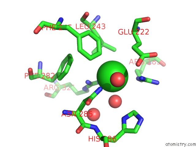
Mono view
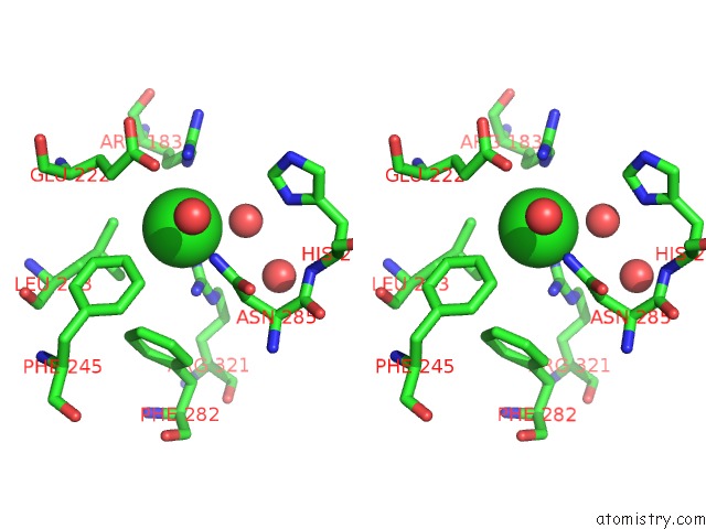
Stereo pair view
Mono view
Stereo pair view
A full contact list of Chlorine with other atoms in the Cl binding
site number 1 of Structure of Tenebrio Molitor Larval Alpha-Amylase within 5.0Å range:
|
Reference:
S.Strobl,
K.Maskos,
M.Betz,
G.Wiegand,
R.Huber,
F.X.Gomis-Ruth,
R.Glockshuber.
Crystal Structure of Yellow Meal Worm Alpha-Amylase at 1.64 A Resolution. J.Mol.Biol. V. 278 617 1998.
ISSN: ISSN 0022-2836
PubMed: 9600843
DOI: 10.1006/JMBI.1998.1667
Page generated: Fri Jul 19 22:56:32 2024
ISSN: ISSN 0022-2836
PubMed: 9600843
DOI: 10.1006/JMBI.1998.1667
Last articles
Zn in 9MJ5Zn in 9HNW
Zn in 9G0L
Zn in 9FNE
Zn in 9DZN
Zn in 9E0I
Zn in 9D32
Zn in 9DAK
Zn in 8ZXC
Zn in 8ZUF