Chlorine »
PDB 1iqj-1jfh »
1jfh »
Chlorine in PDB 1jfh: Structure of A Pancreatic Alpha-Amylase Bound to A Substrate Analogue at 2.03 Angstrom Resolution
Enzymatic activity of Structure of A Pancreatic Alpha-Amylase Bound to A Substrate Analogue at 2.03 Angstrom Resolution
All present enzymatic activity of Structure of A Pancreatic Alpha-Amylase Bound to A Substrate Analogue at 2.03 Angstrom Resolution:
3.2.1.1;
3.2.1.1;
Protein crystallography data
The structure of Structure of A Pancreatic Alpha-Amylase Bound to A Substrate Analogue at 2.03 Angstrom Resolution, PDB code: 1jfh
was solved by
M.Qian,
F.Payan,
with X-Ray Crystallography technique. A brief refinement statistics is given in the table below:
| Resolution Low / High (Å) | 35.00 / 2.03 |
| Space group | P 21 21 21 |
| Cell size a, b, c (Å), α, β, γ (°) | 56.300, 87.800, 103.400, 90.00, 90.00, 90.00 |
| R / Rfree (%) | 16 / 18.5 |
Other elements in 1jfh:
The structure of Structure of A Pancreatic Alpha-Amylase Bound to A Substrate Analogue at 2.03 Angstrom Resolution also contains other interesting chemical elements:
| Mercury | (Hg) | 1 atom |
| Calcium | (Ca) | 1 atom |
Chlorine Binding Sites:
The binding sites of Chlorine atom in the Structure of A Pancreatic Alpha-Amylase Bound to A Substrate Analogue at 2.03 Angstrom Resolution
(pdb code 1jfh). This binding sites where shown within
5.0 Angstroms radius around Chlorine atom.
In total only one binding site of Chlorine was determined in the Structure of A Pancreatic Alpha-Amylase Bound to A Substrate Analogue at 2.03 Angstrom Resolution, PDB code: 1jfh:
In total only one binding site of Chlorine was determined in the Structure of A Pancreatic Alpha-Amylase Bound to A Substrate Analogue at 2.03 Angstrom Resolution, PDB code: 1jfh:
Chlorine binding site 1 out of 1 in 1jfh
Go back to
Chlorine binding site 1 out
of 1 in the Structure of A Pancreatic Alpha-Amylase Bound to A Substrate Analogue at 2.03 Angstrom Resolution
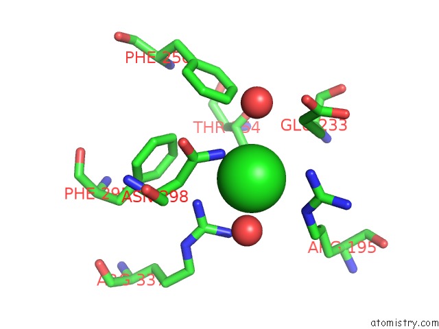
Mono view
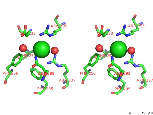
Stereo pair view
Mono view
Stereo pair view
A full contact list of Chlorine with other atoms in the Cl binding
site number 1 of Structure of A Pancreatic Alpha-Amylase Bound to A Substrate Analogue at 2.03 Angstrom Resolution within 5.0Å range:
|
Reference:
M.Qian,
S.Spinelli,
H.Driguez,
F.Payan.
Structure of A Pancreatic Alpha-Amylase Bound to A Substrate Analogue at 2.03 A Resolution. Protein Sci. V. 6 2285 1997.
ISSN: ISSN 0961-8368
PubMed: 9385631
Page generated: Fri Jul 19 22:58:33 2024
ISSN: ISSN 0961-8368
PubMed: 9385631
Last articles
Zn in 9J0NZn in 9J0O
Zn in 9J0P
Zn in 9FJX
Zn in 9EKB
Zn in 9C0F
Zn in 9CAH
Zn in 9CH0
Zn in 9CH3
Zn in 9CH1