Chlorine »
PDB 1jfv-1k73 »
1jom »
Chlorine in PDB 1jom: The Crystal Structure of the Binary Complex Between Folinic Acid (Leucovorin) and E. Coli Dihydrofolate Reductase
Enzymatic activity of The Crystal Structure of the Binary Complex Between Folinic Acid (Leucovorin) and E. Coli Dihydrofolate Reductase
All present enzymatic activity of The Crystal Structure of the Binary Complex Between Folinic Acid (Leucovorin) and E. Coli Dihydrofolate Reductase:
1.5.1.3;
1.5.1.3;
Protein crystallography data
The structure of The Crystal Structure of the Binary Complex Between Folinic Acid (Leucovorin) and E. Coli Dihydrofolate Reductase, PDB code: 1jom
was solved by
V.M.Reyes,
H.Lee,
J.Kraut,
with X-Ray Crystallography technique. A brief refinement statistics is given in the table below:
| Resolution Low / High (Å) | N/A / 1.90 |
| Space group | P 65 |
| Cell size a, b, c (Å), α, β, γ (°) | 96.862, 96.862, 35.022, 90.00, 90.00, 120.00 |
| R / Rfree (%) | 14.2 / n/a |
Other elements in 1jom:
The structure of The Crystal Structure of the Binary Complex Between Folinic Acid (Leucovorin) and E. Coli Dihydrofolate Reductase also contains other interesting chemical elements:
| Calcium | (Ca) | 1 atom |
Chlorine Binding Sites:
The binding sites of Chlorine atom in the The Crystal Structure of the Binary Complex Between Folinic Acid (Leucovorin) and E. Coli Dihydrofolate Reductase
(pdb code 1jom). This binding sites where shown within
5.0 Angstroms radius around Chlorine atom.
In total only one binding site of Chlorine was determined in the The Crystal Structure of the Binary Complex Between Folinic Acid (Leucovorin) and E. Coli Dihydrofolate Reductase, PDB code: 1jom:
In total only one binding site of Chlorine was determined in the The Crystal Structure of the Binary Complex Between Folinic Acid (Leucovorin) and E. Coli Dihydrofolate Reductase, PDB code: 1jom:
Chlorine binding site 1 out of 1 in 1jom
Go back to
Chlorine binding site 1 out
of 1 in the The Crystal Structure of the Binary Complex Between Folinic Acid (Leucovorin) and E. Coli Dihydrofolate Reductase
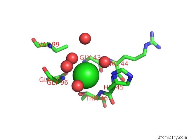
Mono view
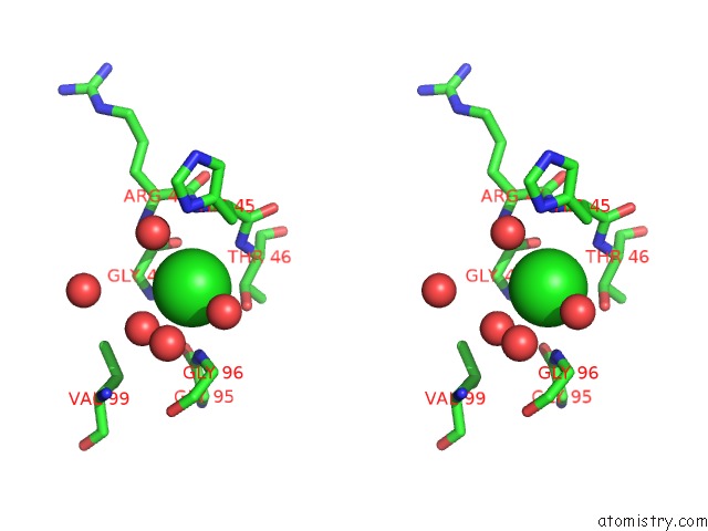
Stereo pair view
Mono view
Stereo pair view
A full contact list of Chlorine with other atoms in the Cl binding
site number 1 of The Crystal Structure of the Binary Complex Between Folinic Acid (Leucovorin) and E. Coli Dihydrofolate Reductase within 5.0Å range:
|
Reference:
H.Lee,
V.M.Reyes,
J.Kraut.
Crystal Structures of Escherichia Coli Dihydrofolate Reductase Complexed with 5-Formyltetrahydrofolate (Folinic Acid) in Two Space Groups: Evidence For Enolization of Pteridine O4. Biochemistry V. 35 7012 1996.
ISSN: ISSN 0006-2960
PubMed: 8679526
DOI: 10.1021/BI960028G
Page generated: Fri Jul 19 23:04:28 2024
ISSN: ISSN 0006-2960
PubMed: 8679526
DOI: 10.1021/BI960028G
Last articles
Zn in 9J0NZn in 9J0O
Zn in 9J0P
Zn in 9FJX
Zn in 9EKB
Zn in 9C0F
Zn in 9CAH
Zn in 9CH0
Zn in 9CH3
Zn in 9CH1