Chlorine »
PDB 1kw5-1l62 »
1kx0 »
Chlorine in PDB 1kx0: Rat Mannose Protein A (H189V I207V) Complexed with Man-A13-Man
Protein crystallography data
The structure of Rat Mannose Protein A (H189V I207V) Complexed with Man-A13-Man, PDB code: 1kx0
was solved by
K.K.Ng,
A.R.Kolatkar,
S.Park-Snyder,
H.Feinberg,
D.A.Clark,
K.Drickamer,
W.I.Weis,
with X-Ray Crystallography technique. A brief refinement statistics is given in the table below:
| Resolution Low / High (Å) | 30.00 / 2.00 |
| Space group | C 1 2 1 |
| Cell size a, b, c (Å), α, β, γ (°) | 79.080, 85.110, 97.660, 90.00, 97.66, 90.00 |
| R / Rfree (%) | 20.1 / 23.2 |
Other elements in 1kx0:
The structure of Rat Mannose Protein A (H189V I207V) Complexed with Man-A13-Man also contains other interesting chemical elements:
| Calcium | (Ca) | 9 atoms |
Chlorine Binding Sites:
The binding sites of Chlorine atom in the Rat Mannose Protein A (H189V I207V) Complexed with Man-A13-Man
(pdb code 1kx0). This binding sites where shown within
5.0 Angstroms radius around Chlorine atom.
In total 3 binding sites of Chlorine where determined in the Rat Mannose Protein A (H189V I207V) Complexed with Man-A13-Man, PDB code: 1kx0:
Jump to Chlorine binding site number: 1; 2; 3;
In total 3 binding sites of Chlorine where determined in the Rat Mannose Protein A (H189V I207V) Complexed with Man-A13-Man, PDB code: 1kx0:
Jump to Chlorine binding site number: 1; 2; 3;
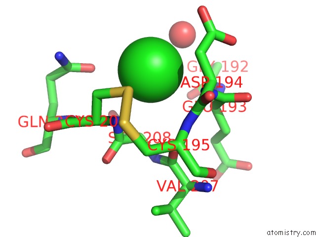
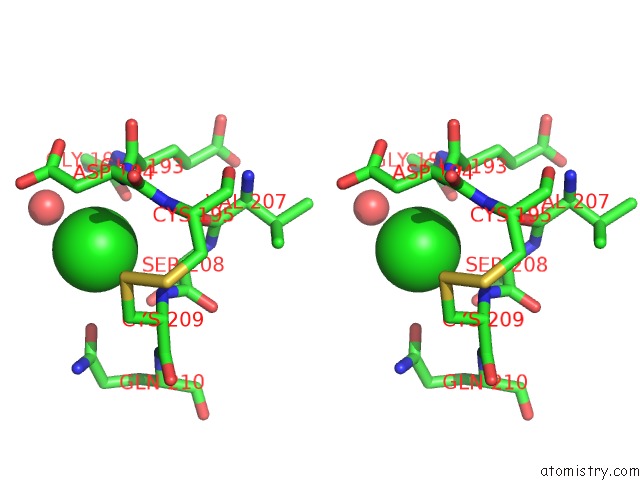
Chlorine binding site 1 out of 3 in 1kx0
Go back to
Chlorine binding site 1 out
of 3 in the Rat Mannose Protein A (H189V I207V) Complexed with Man-A13-Man
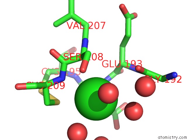
Mono view
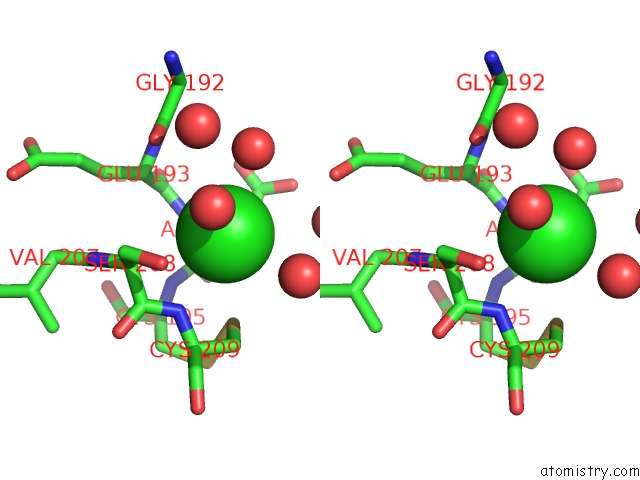
Stereo pair view
Mono view
Stereo pair view
A full contact list of Chlorine with other atoms in the Cl binding
site number 1 of Rat Mannose Protein A (H189V I207V) Complexed with Man-A13-Man within 5.0Å range:
|
Chlorine binding site 2 out of 3 in 1kx0
Go back to
Chlorine binding site 2 out
of 3 in the Rat Mannose Protein A (H189V I207V) Complexed with Man-A13-Man
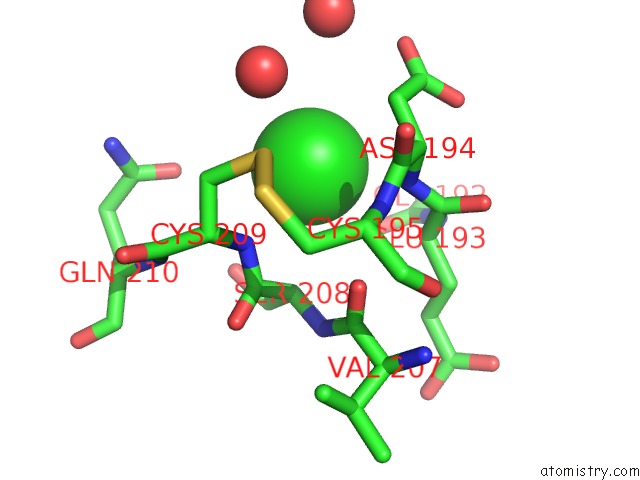
Mono view
Stereo pair view
Mono view
Stereo pair view
A full contact list of Chlorine with other atoms in the Cl binding
site number 2 of Rat Mannose Protein A (H189V I207V) Complexed with Man-A13-Man within 5.0Å range:
|
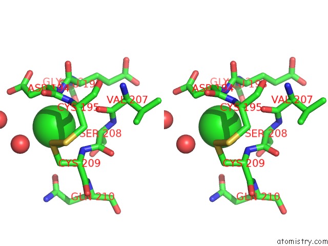
Chlorine binding site 3 out of 3 in 1kx0
Go back to
Chlorine binding site 3 out
of 3 in the Rat Mannose Protein A (H189V I207V) Complexed with Man-A13-Man
Mono view
Stereo pair view
Mono view
Stereo pair view
A full contact list of Chlorine with other atoms in the Cl binding
site number 3 of Rat Mannose Protein A (H189V I207V) Complexed with Man-A13-Man within 5.0Å range:
|
Reference:
K.K.Ng,
A.R.Kolatkar,
S.Park-Snyder,
H.Feinberg,
D.A.Clark,
K.Drickamer,
W.I.Weis.
Orientation of Bound Ligands in Mannose-Binding Proteins. Implications For Multivalent Ligand Recognition. J.Biol.Chem. V. 277 16088 2002.
ISSN: ISSN 0021-9258
PubMed: 11850428
DOI: 10.1074/JBC.M200493200
Page generated: Fri Jul 19 23:30:13 2024
ISSN: ISSN 0021-9258
PubMed: 11850428
DOI: 10.1074/JBC.M200493200
Last articles
Zn in 9J0NZn in 9J0O
Zn in 9J0P
Zn in 9FJX
Zn in 9EKB
Zn in 9C0F
Zn in 9CAH
Zn in 9CH0
Zn in 9CH3
Zn in 9CH1