Chlorine »
PDB 1l63-1l9m »
1l7x »
Chlorine in PDB 1l7x: Human Liver Glycogen Phosphorylase B Complexed with Caffeine, N- Acetyl-Beta-D-Glucopyranosylamine, and Cp-403,700
Enzymatic activity of Human Liver Glycogen Phosphorylase B Complexed with Caffeine, N- Acetyl-Beta-D-Glucopyranosylamine, and Cp-403,700
All present enzymatic activity of Human Liver Glycogen Phosphorylase B Complexed with Caffeine, N- Acetyl-Beta-D-Glucopyranosylamine, and Cp-403,700:
2.4.1.1;
2.4.1.1;
Protein crystallography data
The structure of Human Liver Glycogen Phosphorylase B Complexed with Caffeine, N- Acetyl-Beta-D-Glucopyranosylamine, and Cp-403,700, PDB code: 1l7x
was solved by
J.L.Ekstrom,
T.A.Pauly,
M.D.Carty,
W.C.Soeller,
J.Culp,
D.E.Danley,
D.J.Hoover,
J.L.Treadway,
E.M.Gibbs,
R.J.Fletterick,
Y.S.N.Day,
D.G.Myszka,
V.L.Rath,
with X-Ray Crystallography technique. A brief refinement statistics is given in the table below:
| Resolution Low / High (Å) | 34.39 / 2.30 |
| Space group | P 31 |
| Cell size a, b, c (Å), α, β, γ (°) | 124.072, 124.072, 122.849, 90.00, 90.00, 120.00 |
| R / Rfree (%) | 20.6 / 25.1 |
Chlorine Binding Sites:
The binding sites of Chlorine atom in the Human Liver Glycogen Phosphorylase B Complexed with Caffeine, N- Acetyl-Beta-D-Glucopyranosylamine, and Cp-403,700
(pdb code 1l7x). This binding sites where shown within
5.0 Angstroms radius around Chlorine atom.
In total 2 binding sites of Chlorine where determined in the Human Liver Glycogen Phosphorylase B Complexed with Caffeine, N- Acetyl-Beta-D-Glucopyranosylamine, and Cp-403,700, PDB code: 1l7x:
Jump to Chlorine binding site number: 1; 2;
In total 2 binding sites of Chlorine where determined in the Human Liver Glycogen Phosphorylase B Complexed with Caffeine, N- Acetyl-Beta-D-Glucopyranosylamine, and Cp-403,700, PDB code: 1l7x:
Jump to Chlorine binding site number: 1; 2;
Chlorine binding site 1 out of 2 in 1l7x
Go back to
Chlorine binding site 1 out
of 2 in the Human Liver Glycogen Phosphorylase B Complexed with Caffeine, N- Acetyl-Beta-D-Glucopyranosylamine, and Cp-403,700
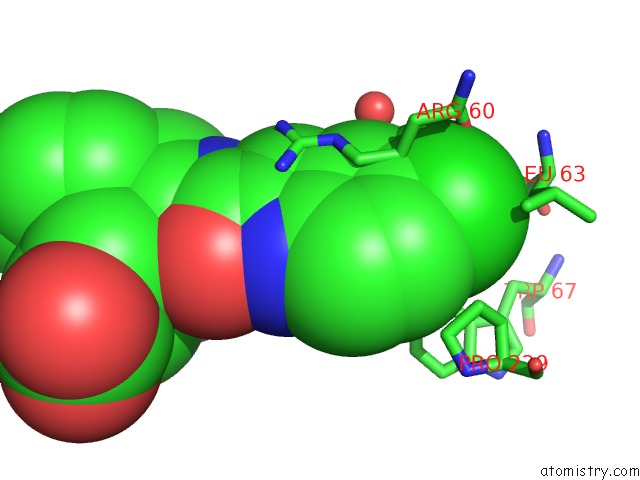
Mono view
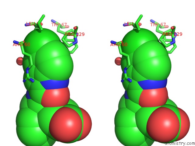
Stereo pair view
Mono view
Stereo pair view
A full contact list of Chlorine with other atoms in the Cl binding
site number 1 of Human Liver Glycogen Phosphorylase B Complexed with Caffeine, N- Acetyl-Beta-D-Glucopyranosylamine, and Cp-403,700 within 5.0Å range:
|
Chlorine binding site 2 out of 2 in 1l7x
Go back to
Chlorine binding site 2 out
of 2 in the Human Liver Glycogen Phosphorylase B Complexed with Caffeine, N- Acetyl-Beta-D-Glucopyranosylamine, and Cp-403,700
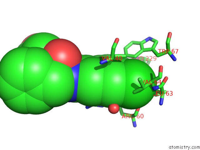
Mono view
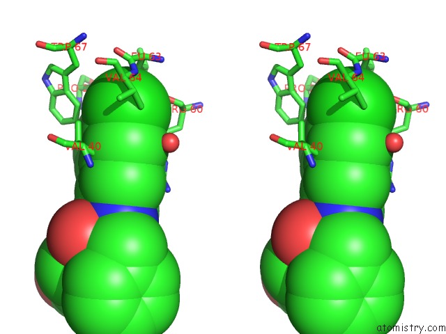
Stereo pair view
Mono view
Stereo pair view
A full contact list of Chlorine with other atoms in the Cl binding
site number 2 of Human Liver Glycogen Phosphorylase B Complexed with Caffeine, N- Acetyl-Beta-D-Glucopyranosylamine, and Cp-403,700 within 5.0Å range:
|
Reference:
J.L.Ekstrom,
T.A.Pauly,
M.D.Carty,
W.C.Soeller,
J.Culp,
D.E.Danley,
D.J.Hoover,
J.L.Treadway,
E.M.Gibbs,
R.J.Fletterick,
Y.S.Day,
D.G.Myszka,
V.L.Rath.
Structure-Activity Analysis of the Purine Binding Site of Human Liver Glycogen Phosphorylase. Chem.Biol. V. 9 915 2002.
ISSN: ISSN 1074-5521
PubMed: 12204691
DOI: 10.1016/S1074-5521(02)00186-2
Page generated: Fri Jul 19 23:41:38 2024
ISSN: ISSN 1074-5521
PubMed: 12204691
DOI: 10.1016/S1074-5521(02)00186-2
Last articles
Zn in 9MJ5Zn in 9HNW
Zn in 9G0L
Zn in 9FNE
Zn in 9DZN
Zn in 9E0I
Zn in 9D32
Zn in 9DAK
Zn in 8ZXC
Zn in 8ZUF