Chlorine »
PDB 1l9n-1lwg »
1lkd »
Chlorine in PDB 1lkd: Crystal Structure of 2,3-Dihydroxybiphenyl 1,2-Dioxygenase (Dhbd) Complexed with 2',6'-Dicl Dihydroxybiphenyl (Dhb)
Enzymatic activity of Crystal Structure of 2,3-Dihydroxybiphenyl 1,2-Dioxygenase (Dhbd) Complexed with 2',6'-Dicl Dihydroxybiphenyl (Dhb)
All present enzymatic activity of Crystal Structure of 2,3-Dihydroxybiphenyl 1,2-Dioxygenase (Dhbd) Complexed with 2',6'-Dicl Dihydroxybiphenyl (Dhb):
1.13.11.39;
1.13.11.39;
Protein crystallography data
The structure of Crystal Structure of 2,3-Dihydroxybiphenyl 1,2-Dioxygenase (Dhbd) Complexed with 2',6'-Dicl Dihydroxybiphenyl (Dhb), PDB code: 1lkd
was solved by
S.Dai,
J.T.Bolin,
with X-Ray Crystallography technique. A brief refinement statistics is given in the table below:
| Resolution Low / High (Å) | 19.97 / 1.70 |
| Space group | I 4 2 2 |
| Cell size a, b, c (Å), α, β, γ (°) | 122.640, 122.640, 107.232, 90.00, 90.00, 90.00 |
| R / Rfree (%) | 20.3 / 22 |
Other elements in 1lkd:
The structure of Crystal Structure of 2,3-Dihydroxybiphenyl 1,2-Dioxygenase (Dhbd) Complexed with 2',6'-Dicl Dihydroxybiphenyl (Dhb) also contains other interesting chemical elements:
| Iron | (Fe) | 2 atoms |
Chlorine Binding Sites:
The binding sites of Chlorine atom in the Crystal Structure of 2,3-Dihydroxybiphenyl 1,2-Dioxygenase (Dhbd) Complexed with 2',6'-Dicl Dihydroxybiphenyl (Dhb)
(pdb code 1lkd). This binding sites where shown within
5.0 Angstroms radius around Chlorine atom.
In total 4 binding sites of Chlorine where determined in the Crystal Structure of 2,3-Dihydroxybiphenyl 1,2-Dioxygenase (Dhbd) Complexed with 2',6'-Dicl Dihydroxybiphenyl (Dhb), PDB code: 1lkd:
Jump to Chlorine binding site number: 1; 2; 3; 4;
In total 4 binding sites of Chlorine where determined in the Crystal Structure of 2,3-Dihydroxybiphenyl 1,2-Dioxygenase (Dhbd) Complexed with 2',6'-Dicl Dihydroxybiphenyl (Dhb), PDB code: 1lkd:
Jump to Chlorine binding site number: 1; 2; 3; 4;
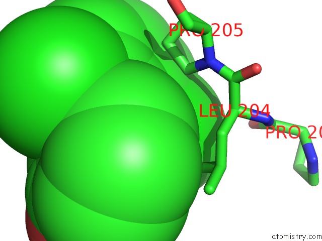
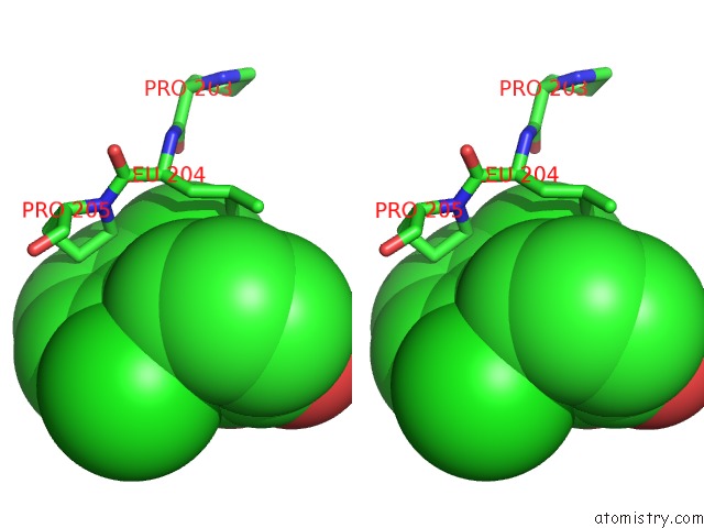
Chlorine binding site 1 out of 4 in 1lkd
Go back to
Chlorine binding site 1 out
of 4 in the Crystal Structure of 2,3-Dihydroxybiphenyl 1,2-Dioxygenase (Dhbd) Complexed with 2',6'-Dicl Dihydroxybiphenyl (Dhb)
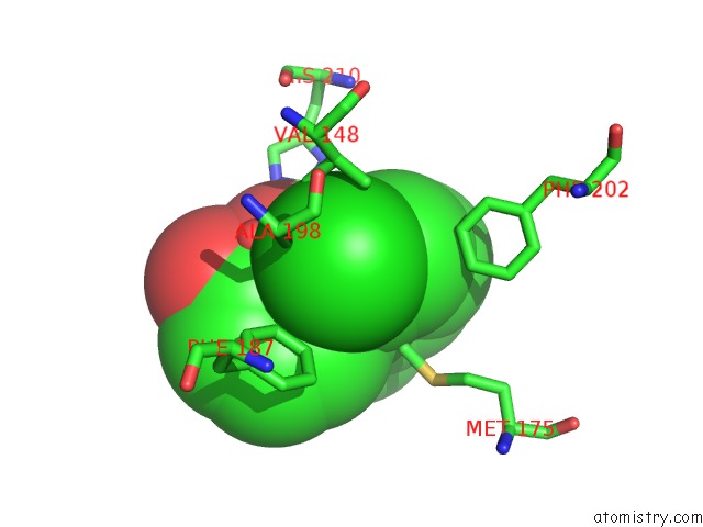
Mono view
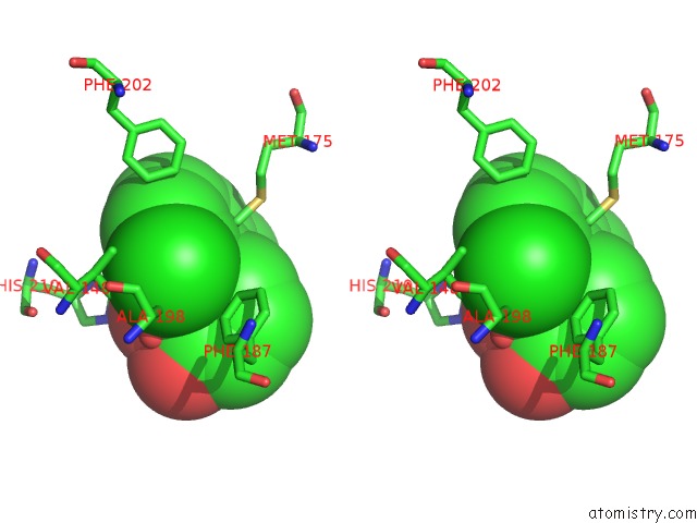
Stereo pair view
Mono view
Stereo pair view
A full contact list of Chlorine with other atoms in the Cl binding
site number 1 of Crystal Structure of 2,3-Dihydroxybiphenyl 1,2-Dioxygenase (Dhbd) Complexed with 2',6'-Dicl Dihydroxybiphenyl (Dhb) within 5.0Å range:
|
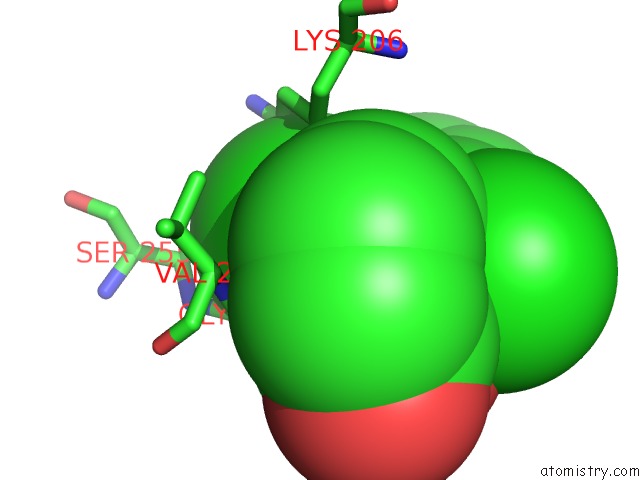
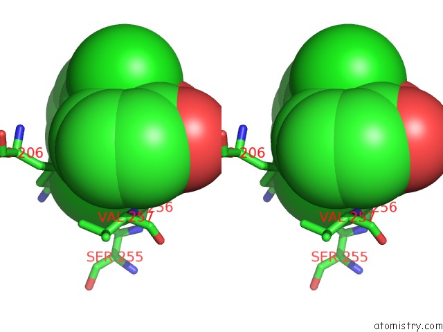
Chlorine binding site 2 out of 4 in 1lkd
Go back to
Chlorine binding site 2 out
of 4 in the Crystal Structure of 2,3-Dihydroxybiphenyl 1,2-Dioxygenase (Dhbd) Complexed with 2',6'-Dicl Dihydroxybiphenyl (Dhb)
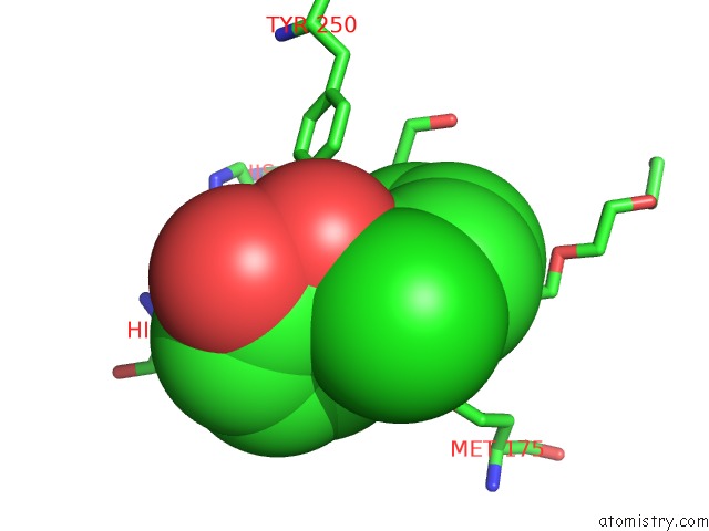
Mono view
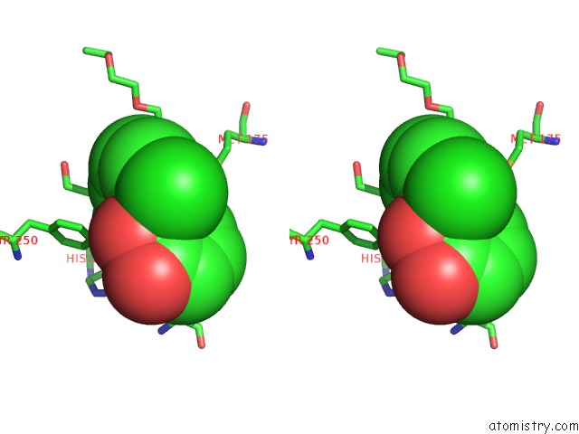
Stereo pair view
Mono view
Stereo pair view
A full contact list of Chlorine with other atoms in the Cl binding
site number 2 of Crystal Structure of 2,3-Dihydroxybiphenyl 1,2-Dioxygenase (Dhbd) Complexed with 2',6'-Dicl Dihydroxybiphenyl (Dhb) within 5.0Å range:
|
Chlorine binding site 3 out of 4 in 1lkd
Go back to
Chlorine binding site 3 out
of 4 in the Crystal Structure of 2,3-Dihydroxybiphenyl 1,2-Dioxygenase (Dhbd) Complexed with 2',6'-Dicl Dihydroxybiphenyl (Dhb)
Mono view
Stereo pair view
Mono view
Stereo pair view
A full contact list of Chlorine with other atoms in the Cl binding
site number 3 of Crystal Structure of 2,3-Dihydroxybiphenyl 1,2-Dioxygenase (Dhbd) Complexed with 2',6'-Dicl Dihydroxybiphenyl (Dhb) within 5.0Å range:
|
Chlorine binding site 4 out of 4 in 1lkd
Go back to
Chlorine binding site 4 out
of 4 in the Crystal Structure of 2,3-Dihydroxybiphenyl 1,2-Dioxygenase (Dhbd) Complexed with 2',6'-Dicl Dihydroxybiphenyl (Dhb)
Mono view
Stereo pair view
Mono view
Stereo pair view
A full contact list of Chlorine with other atoms in the Cl binding
site number 4 of Crystal Structure of 2,3-Dihydroxybiphenyl 1,2-Dioxygenase (Dhbd) Complexed with 2',6'-Dicl Dihydroxybiphenyl (Dhb) within 5.0Å range:
|
Reference:
S.Dai,
F.H.Vaillancourt,
H.Maaroufi,
N.M.Drouin,
D.B.Neau,
V.Snieckus,
J.T.Bolin,
L.D.Eltis.
Identification and Analysis of A Bottleneck in Pcb Biodegradation Nat.Struct.Biol. V. 9 934 2002.
ISSN: ISSN 1072-8368
PubMed: 12415290
DOI: 10.1038/NSB866
Page generated: Fri Jul 19 23:53:16 2024
ISSN: ISSN 1072-8368
PubMed: 12415290
DOI: 10.1038/NSB866
Last articles
Zn in 9MJ5Zn in 9HNW
Zn in 9G0L
Zn in 9FNE
Zn in 9DZN
Zn in 9E0I
Zn in 9D32
Zn in 9DAK
Zn in 8ZXC
Zn in 8ZUF