Chlorine »
PDB 1l9n-1lwg »
1ltz »
Chlorine in PDB 1ltz: Crystal Structure of Chromobacterium Violaceum Phenylalanine Hydroxylase, Structure Has Bound Iron (III) and Oxidized Cofactor 7, 8-Dihydrobiopterin
Enzymatic activity of Crystal Structure of Chromobacterium Violaceum Phenylalanine Hydroxylase, Structure Has Bound Iron (III) and Oxidized Cofactor 7, 8-Dihydrobiopterin
All present enzymatic activity of Crystal Structure of Chromobacterium Violaceum Phenylalanine Hydroxylase, Structure Has Bound Iron (III) and Oxidized Cofactor 7, 8-Dihydrobiopterin:
1.14.16.1;
1.14.16.1;
Protein crystallography data
The structure of Crystal Structure of Chromobacterium Violaceum Phenylalanine Hydroxylase, Structure Has Bound Iron (III) and Oxidized Cofactor 7, 8-Dihydrobiopterin, PDB code: 1ltz
was solved by
H.Erlandsen,
J.Y.Kim,
M.G.Patch,
A.Han,
A.Volner,
M.M.Abu-Omar,
R.C.Stevens,
with X-Ray Crystallography technique. A brief refinement statistics is given in the table below:
| Resolution Low / High (Å) | 20.00 / 1.40 |
| Space group | P 21 21 21 |
| Cell size a, b, c (Å), α, β, γ (°) | 46.700, 67.860, 91.470, 90.00, 90.00, 90.00 |
| R / Rfree (%) | 15.9 / 22.3 |
Other elements in 1ltz:
The structure of Crystal Structure of Chromobacterium Violaceum Phenylalanine Hydroxylase, Structure Has Bound Iron (III) and Oxidized Cofactor 7, 8-Dihydrobiopterin also contains other interesting chemical elements:
| Iron | (Fe) | 1 atom |
Chlorine Binding Sites:
The binding sites of Chlorine atom in the Crystal Structure of Chromobacterium Violaceum Phenylalanine Hydroxylase, Structure Has Bound Iron (III) and Oxidized Cofactor 7, 8-Dihydrobiopterin
(pdb code 1ltz). This binding sites where shown within
5.0 Angstroms radius around Chlorine atom.
In total only one binding site of Chlorine was determined in the Crystal Structure of Chromobacterium Violaceum Phenylalanine Hydroxylase, Structure Has Bound Iron (III) and Oxidized Cofactor 7, 8-Dihydrobiopterin, PDB code: 1ltz:
In total only one binding site of Chlorine was determined in the Crystal Structure of Chromobacterium Violaceum Phenylalanine Hydroxylase, Structure Has Bound Iron (III) and Oxidized Cofactor 7, 8-Dihydrobiopterin, PDB code: 1ltz:
Chlorine binding site 1 out of 1 in 1ltz
Go back to
Chlorine binding site 1 out
of 1 in the Crystal Structure of Chromobacterium Violaceum Phenylalanine Hydroxylase, Structure Has Bound Iron (III) and Oxidized Cofactor 7, 8-Dihydrobiopterin
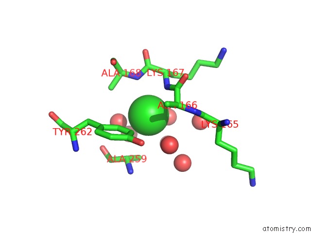
Mono view
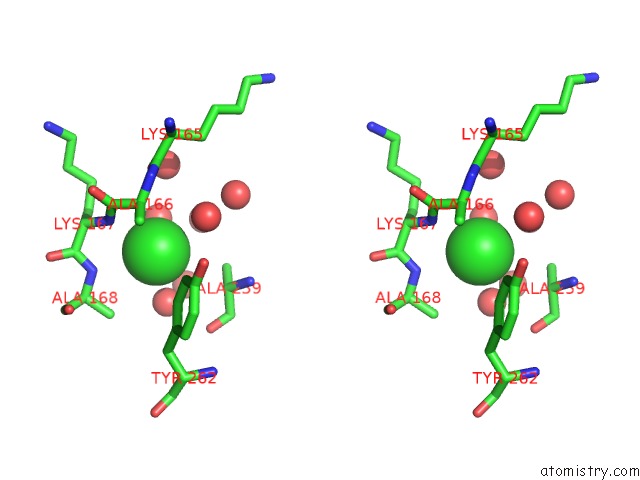
Stereo pair view
Mono view
Stereo pair view
A full contact list of Chlorine with other atoms in the Cl binding
site number 1 of Crystal Structure of Chromobacterium Violaceum Phenylalanine Hydroxylase, Structure Has Bound Iron (III) and Oxidized Cofactor 7, 8-Dihydrobiopterin within 5.0Å range:
|
Reference:
H.Erlandsen,
J.Y.Kim,
M.G.Patch,
A.Han,
A.Volner,
M.M.Abu-Omar,
R.C.Stevens.
Structural Comparison of Bacterial and Human Iron-Dependent Phenylalanine Hydroxylases: Similar Fold, Different Stability and Reaction Rates. J.Mol.Biol. V. 320 645 2002.
ISSN: ISSN 0022-2836
PubMed: 12096915
DOI: 10.1016/S0022-2836(02)00496-5
Page generated: Fri Jul 19 23:55:31 2024
ISSN: ISSN 0022-2836
PubMed: 12096915
DOI: 10.1016/S0022-2836(02)00496-5
Last articles
Zn in 9J0NZn in 9J0O
Zn in 9J0P
Zn in 9FJX
Zn in 9EKB
Zn in 9C0F
Zn in 9CAH
Zn in 9CH0
Zn in 9CH3
Zn in 9CH1