Chlorine »
PDB 1mkp-1nc4 »
1mx5 »
Chlorine in PDB 1mx5: Crystal Structure of Human Liver Carboxylesterase in Complexed with Homatropine, A Cocaine Analogue
Enzymatic activity of Crystal Structure of Human Liver Carboxylesterase in Complexed with Homatropine, A Cocaine Analogue
All present enzymatic activity of Crystal Structure of Human Liver Carboxylesterase in Complexed with Homatropine, A Cocaine Analogue:
3.1.1.1;
3.1.1.1;
Protein crystallography data
The structure of Crystal Structure of Human Liver Carboxylesterase in Complexed with Homatropine, A Cocaine Analogue, PDB code: 1mx5
was solved by
S.Bencharit,
C.L.Morton,
Y.Xue,
P.M.Potter,
M.R.Redinbo,
with X-Ray Crystallography technique. A brief refinement statistics is given in the table below:
| Resolution Low / High (Å) | 19.96 / 2.80 |
| Space group | P 1 21 1 |
| Cell size a, b, c (Å), α, β, γ (°) | 55.400, 178.800, 199.600, 90.00, 90.20, 90.00 |
| R / Rfree (%) | 15.8 / 22.1 |
Chlorine Binding Sites:
The binding sites of Chlorine atom in the Crystal Structure of Human Liver Carboxylesterase in Complexed with Homatropine, A Cocaine Analogue
(pdb code 1mx5). This binding sites where shown within
5.0 Angstroms radius around Chlorine atom.
In total 2 binding sites of Chlorine where determined in the Crystal Structure of Human Liver Carboxylesterase in Complexed with Homatropine, A Cocaine Analogue, PDB code: 1mx5:
Jump to Chlorine binding site number: 1; 2;
In total 2 binding sites of Chlorine where determined in the Crystal Structure of Human Liver Carboxylesterase in Complexed with Homatropine, A Cocaine Analogue, PDB code: 1mx5:
Jump to Chlorine binding site number: 1; 2;
Chlorine binding site 1 out of 2 in 1mx5
Go back to
Chlorine binding site 1 out
of 2 in the Crystal Structure of Human Liver Carboxylesterase in Complexed with Homatropine, A Cocaine Analogue
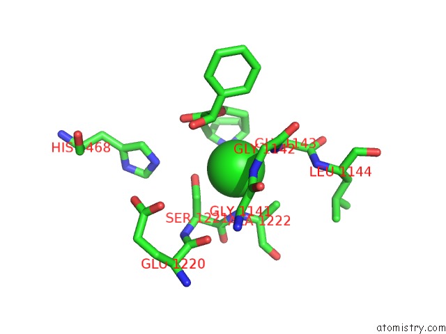
Mono view
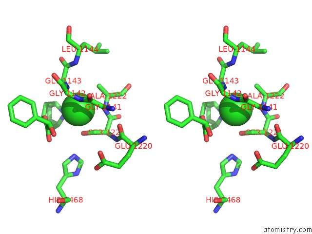
Stereo pair view
Mono view
Stereo pair view
A full contact list of Chlorine with other atoms in the Cl binding
site number 1 of Crystal Structure of Human Liver Carboxylesterase in Complexed with Homatropine, A Cocaine Analogue within 5.0Å range:
|
Chlorine binding site 2 out of 2 in 1mx5
Go back to
Chlorine binding site 2 out
of 2 in the Crystal Structure of Human Liver Carboxylesterase in Complexed with Homatropine, A Cocaine Analogue
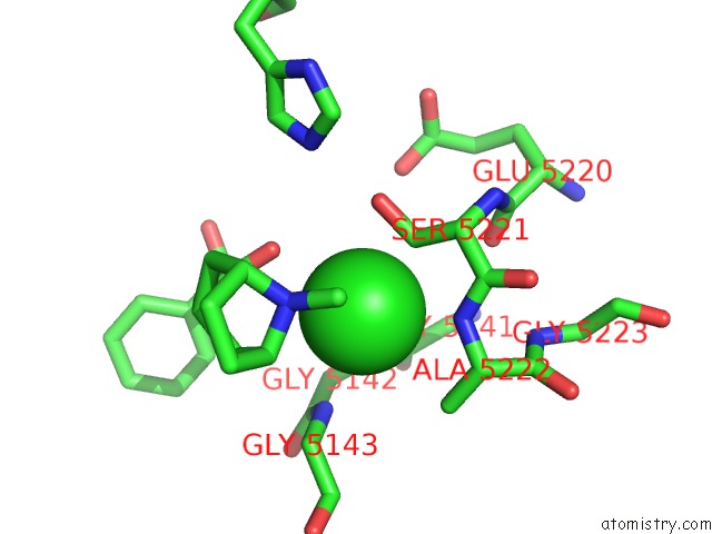
Mono view
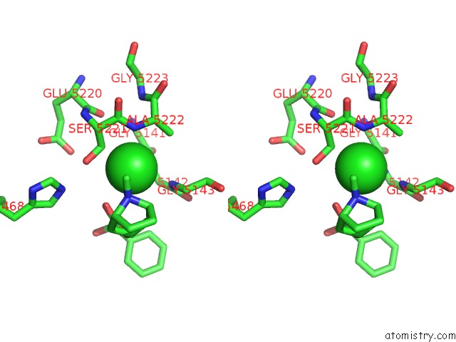
Stereo pair view
Mono view
Stereo pair view
A full contact list of Chlorine with other atoms in the Cl binding
site number 2 of Crystal Structure of Human Liver Carboxylesterase in Complexed with Homatropine, A Cocaine Analogue within 5.0Å range:
|
Reference:
S.Bencharit,
C.L.Morton,
Y.Xue,
P.M.Potter,
M.R.Redinbo.
Structural Basis of Heroin and Cocaine Metabolism By A Promiscuous Human Drug-Processing Enzyme Nat.Struct.Biol. V. 10 349 2003.
ISSN: ISSN 1072-8368
PubMed: 12679808
DOI: 10.1038/NSB919
Page generated: Sat Jul 20 00:14:51 2024
ISSN: ISSN 1072-8368
PubMed: 12679808
DOI: 10.1038/NSB919
Last articles
Zn in 9J0NZn in 9J0O
Zn in 9J0P
Zn in 9FJX
Zn in 9EKB
Zn in 9C0F
Zn in 9CAH
Zn in 9CH0
Zn in 9CH3
Zn in 9CH1