Chlorine »
PDB 1nc7-1nvf »
1nnl »
Chlorine in PDB 1nnl: Crystal Structure of Human Phosphoserine Phosphatase
Enzymatic activity of Crystal Structure of Human Phosphoserine Phosphatase
All present enzymatic activity of Crystal Structure of Human Phosphoserine Phosphatase:
3.1.3.3;
3.1.3.3;
Protein crystallography data
The structure of Crystal Structure of Human Phosphoserine Phosphatase, PDB code: 1nnl
was solved by
Y.Peeraer,
A.Rabijns,
C.Verboven,
J.F.Collet,
E.Van Schaftingen,
C.De Ranter,
with X-Ray Crystallography technique. A brief refinement statistics is given in the table below:
| Resolution Low / High (Å) | 19.86 / 1.53 |
| Space group | C 2 2 21 |
| Cell size a, b, c (Å), α, β, γ (°) | 49.033, 130.249, 157.293, 90.00, 90.00, 90.00 |
| R / Rfree (%) | 21.7 / 23.4 |
Other elements in 1nnl:
The structure of Crystal Structure of Human Phosphoserine Phosphatase also contains other interesting chemical elements:
| Calcium | (Ca) | 3 atoms |
Chlorine Binding Sites:
The binding sites of Chlorine atom in the Crystal Structure of Human Phosphoserine Phosphatase
(pdb code 1nnl). This binding sites where shown within
5.0 Angstroms radius around Chlorine atom.
In total 6 binding sites of Chlorine where determined in the Crystal Structure of Human Phosphoserine Phosphatase, PDB code: 1nnl:
Jump to Chlorine binding site number: 1; 2; 3; 4; 5; 6;
In total 6 binding sites of Chlorine where determined in the Crystal Structure of Human Phosphoserine Phosphatase, PDB code: 1nnl:
Jump to Chlorine binding site number: 1; 2; 3; 4; 5; 6;
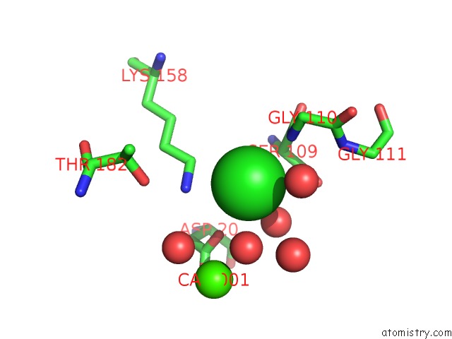
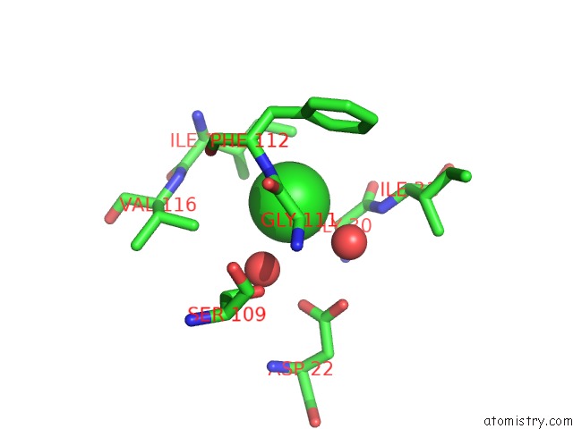
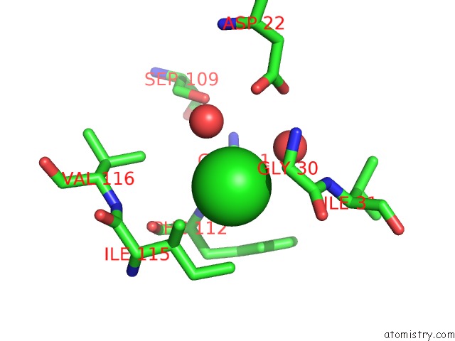
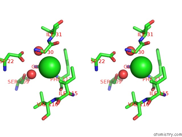
Chlorine binding site 1 out of 6 in 1nnl
Go back to
Chlorine binding site 1 out
of 6 in the Crystal Structure of Human Phosphoserine Phosphatase
Mono view
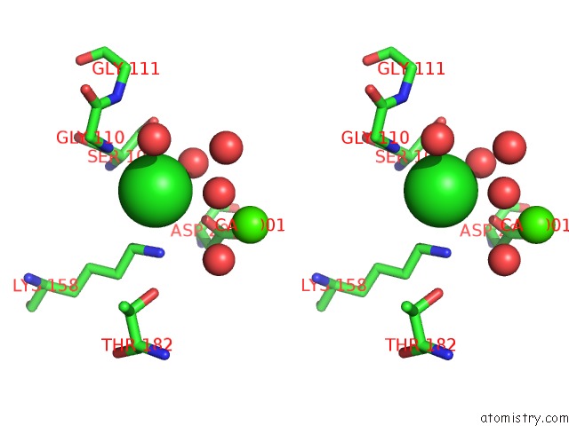
Stereo pair view
Mono view
Stereo pair view
A full contact list of Chlorine with other atoms in the Cl binding
site number 1 of Crystal Structure of Human Phosphoserine Phosphatase within 5.0Å range:
|
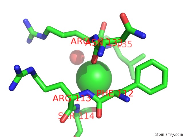
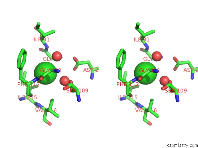
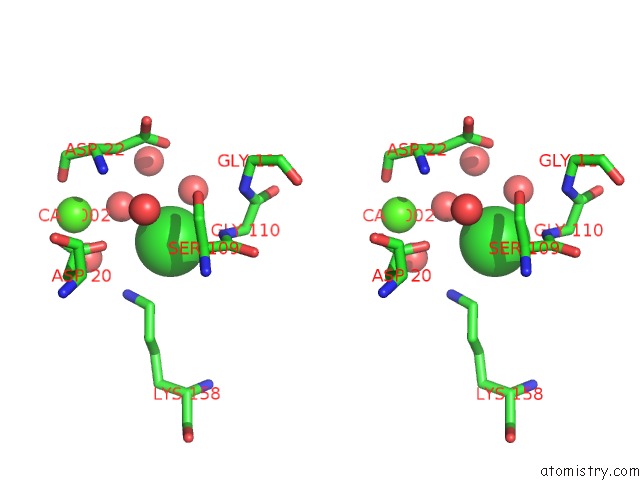
Chlorine binding site 2 out of 6 in 1nnl
Go back to
Chlorine binding site 2 out
of 6 in the Crystal Structure of Human Phosphoserine Phosphatase
Mono view
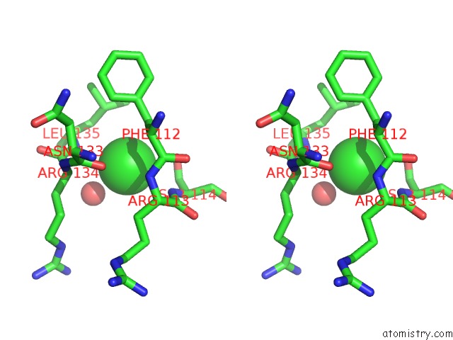
Stereo pair view
Mono view
Stereo pair view
A full contact list of Chlorine with other atoms in the Cl binding
site number 2 of Crystal Structure of Human Phosphoserine Phosphatase within 5.0Å range:
|
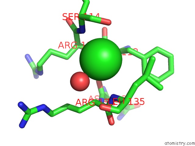
Chlorine binding site 3 out of 6 in 1nnl
Go back to
Chlorine binding site 3 out
of 6 in the Crystal Structure of Human Phosphoserine Phosphatase
Mono view
Stereo pair view
Mono view
Stereo pair view
A full contact list of Chlorine with other atoms in the Cl binding
site number 3 of Crystal Structure of Human Phosphoserine Phosphatase within 5.0Å range:
|
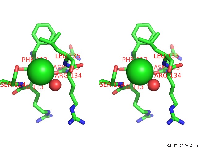
Chlorine binding site 4 out of 6 in 1nnl
Go back to
Chlorine binding site 4 out
of 6 in the Crystal Structure of Human Phosphoserine Phosphatase
Mono view
Stereo pair view
Mono view
Stereo pair view
A full contact list of Chlorine with other atoms in the Cl binding
site number 4 of Crystal Structure of Human Phosphoserine Phosphatase within 5.0Å range:
|
Chlorine binding site 5 out of 6 in 1nnl
Go back to
Chlorine binding site 5 out
of 6 in the Crystal Structure of Human Phosphoserine Phosphatase
Mono view
Stereo pair view
Mono view
Stereo pair view
A full contact list of Chlorine with other atoms in the Cl binding
site number 5 of Crystal Structure of Human Phosphoserine Phosphatase within 5.0Å range:
|
Chlorine binding site 6 out of 6 in 1nnl
Go back to
Chlorine binding site 6 out
of 6 in the Crystal Structure of Human Phosphoserine Phosphatase

Mono view
Stereo pair view
Mono view
Stereo pair view
A full contact list of Chlorine with other atoms in the Cl binding
site number 6 of Crystal Structure of Human Phosphoserine Phosphatase within 5.0Å range:
|
Reference:
Y.Peeraer,
A.Rabijns,
C.Verboven,
J.F.Collet,
E.Van Schaftingen,
C.De Ranter.
High-Resolution Structure of Human Phosphoserine Phosphatase in Open Conformation. Acta Crystallogr.,Sect.D V. 59 971 2003.
ISSN: ISSN 0907-4449
PubMed: 12777757
DOI: 10.1107/S0907444903005407
Page generated: Sat Jul 20 00:34:20 2024
ISSN: ISSN 0907-4449
PubMed: 12777757
DOI: 10.1107/S0907444903005407
Last articles
Zn in 9J0NZn in 9J0O
Zn in 9J0P
Zn in 9FJX
Zn in 9EKB
Zn in 9C0F
Zn in 9CAH
Zn in 9CH0
Zn in 9CH3
Zn in 9CH1