Chlorine in PDB 1nsl: Crystal Structure of Probable Acetyltransferase
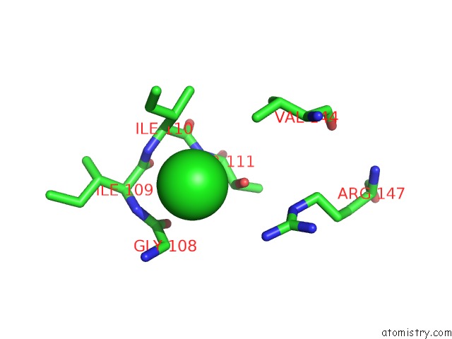
Protein crystallography data
The structure of Crystal Structure of Probable Acetyltransferase, PDB code: 1nsl
was solved by
J.S.Brunzelle,
S.V.Korolev,
R.Wu,
A.Joachimiak,
W.F.Anderson,
Midwestcenter For Structural Genomics (Mcsg),
with X-Ray Crystallography technique. A brief refinement statistics is given in the table below:
Resolution Low / High (Å)
|
20.00 /
2.70
|
Space group
|
P 1 21 1
|
Cell size a, b, c (Å), α, β, γ (°)
|
59.339,
134.185,
91.062,
90.00,
104.08,
90.00
|
R / Rfree (%)
|
25.6 /
27.8
|
Chlorine Binding Sites:
The binding sites of Chlorine atom in the Crystal Structure of Probable Acetyltransferase
(pdb code 1nsl). This binding sites where shown within
5.0 Angstroms radius around Chlorine atom.
In total 6 binding sites of Chlorine where determined in the
Crystal Structure of Probable Acetyltransferase, PDB code: 1nsl:
Jump to Chlorine binding site number:
1;
2;
3;
4;
5;
6;
Chlorine binding site 1 out
of 6 in 1nsl
Go back to
Chlorine Binding Sites List in 1nsl
Chlorine binding site 1 out
of 6 in the Crystal Structure of Probable Acetyltransferase
 Mono view
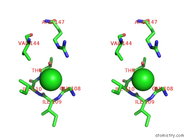
 Stereo pair view
|
A full contact list of Chlorine with other atoms in the Cl binding
site number 1 of Crystal Structure of Probable Acetyltransferase within 5.0Å range:
probe
|
atom
|
residue
|
distance (Å)
|
B
|
Occ
|
A:Cl184
b:82.4
occ:1.00
|
OG1
|
A:THR111
|
3.5
|
27.9
|
1.0
|
N
|
A:THR111
|
3.7
|
28.1
|
1.0
|
CA
|
A:GLY108
|
3.7
|
32.2
|
1.0
|
N
|
A:ILE110
|
3.7
|
31.3
|
1.0
|
C
|
A:GLY108
|
3.8
|
33.0
|
1.0
|
N
|
A:ILE109
|
3.8
|
32.7
|
1.0
|
CB
|
A:ILE110
|
3.8
|
28.4
|
1.0
|
CA
|
A:ILE110
|
4.2
|
29.3
|
1.0
|
N
|
A:GLY108
|
4.3
|
32.1
|
1.0
|
CB
|
A:THR111
|
4.3
|
27.5
|
1.0
|
CG1
|
A:ILE110
|
4.4
|
26.9
|
1.0
|
C
|
A:ILE109
|
4.4
|
32.6
|
1.0
|
NH2
|
A:ARG147
|
4.4
|
38.8
|
1.0
|
O
|
A:GLY108
|
4.4
|
33.2
|
1.0
|
C
|
A:ILE110
|
4.4
|
29.3
|
1.0
|
CG2
|
A:VAL144
|
4.6
|
26.4
|
1.0
|
CA
|
A:THR111
|
4.6
|
27.7
|
1.0
|
CA
|
A:ILE109
|
4.6
|
32.8
|
1.0
|
CD1
|
A:ILE110
|
4.9
|
18.2
|
1.0
|
CG2
|
A:ILE110
|
5.0
|
30.6
|
1.0
|
|
Chlorine binding site 2 out
of 6 in 1nsl
Go back to
Chlorine Binding Sites List in 1nsl
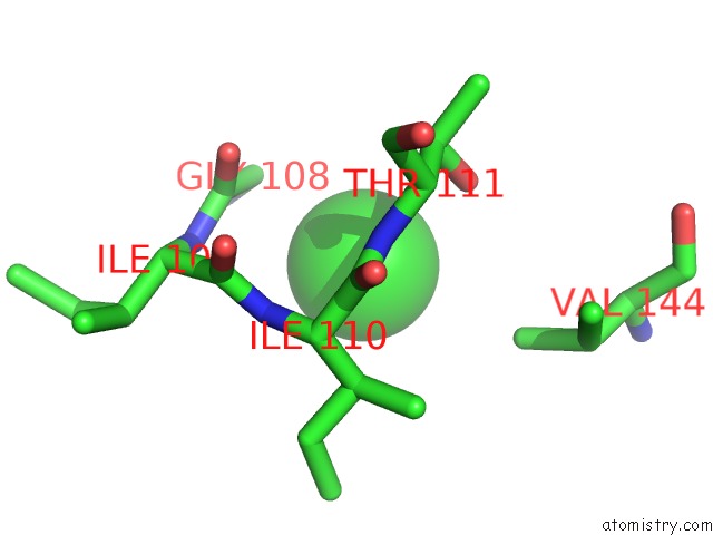
Chlorine binding site 2 out
of 6 in the Crystal Structure of Probable Acetyltransferase
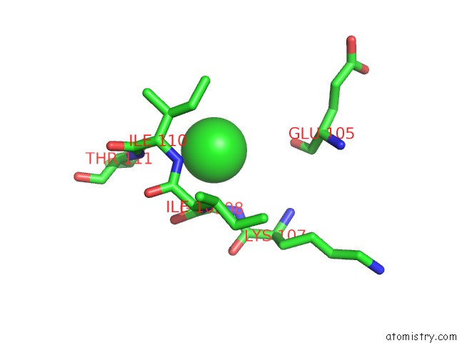
 Mono view
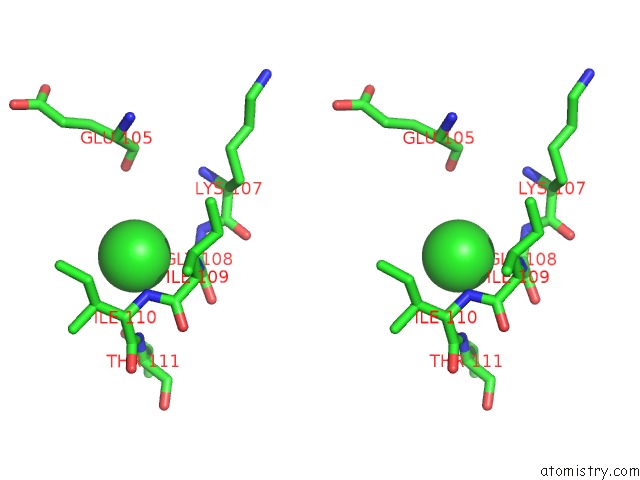
 Stereo pair view
|
A full contact list of Chlorine with other atoms in the Cl binding
site number 2 of Crystal Structure of Probable Acetyltransferase within 5.0Å range:
probe
|
atom
|
residue
|
distance (Å)
|
B
|
Occ
|
B:Cl184
b:80.4
occ:1.00
|
CA
|
B:GLY108
|
3.2
|
33.0
|
1.0
|
N
|
B:ILE109
|
3.3
|
32.2
|
1.0
|
C
|
B:GLY108
|
3.4
|
32.5
|
1.0
|
N
|
B:ILE110
|
3.4
|
31.6
|
1.0
|
N
|
B:GLY108
|
3.6
|
33.5
|
1.0
|
CB
|
B:ILE110
|
3.9
|
31.1
|
1.0
|
N
|
B:THR111
|
4.0
|
29.9
|
1.0
|
C
|
B:ILE109
|
4.1
|
32.1
|
1.0
|
CA
|
B:ILE110
|
4.1
|
31.2
|
1.0
|
CA
|
B:ILE109
|
4.2
|
32.3
|
1.0
|
O
|
B:GLY108
|
4.2
|
32.2
|
1.0
|
CG1
|
B:ILE110
|
4.2
|
30.0
|
1.0
|
OG1
|
B:THR111
|
4.3
|
27.1
|
1.0
|
O
|
B:GLU105
|
4.4
|
28.8
|
1.0
|
C
|
B:ILE110
|
4.6
|
31.0
|
1.0
|
CD1
|
B:ILE110
|
4.6
|
30.6
|
1.0
|
C
|
B:LYS107
|
4.7
|
33.4
|
1.0
|
CB
|
B:ILE109
|
4.8
|
31.9
|
1.0
|
CB
|
B:THR111
|
4.8
|
29.3
|
1.0
|
|
Chlorine binding site 3 out
of 6 in 1nsl
Go back to
Chlorine Binding Sites List in 1nsl
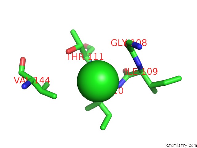
Chlorine binding site 3 out
of 6 in the Crystal Structure of Probable Acetyltransferase
 Mono view
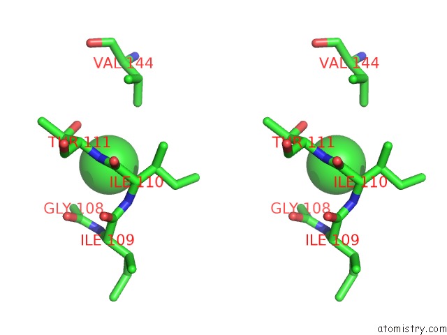
 Stereo pair view
|
A full contact list of Chlorine with other atoms in the Cl binding
site number 3 of Crystal Structure of Probable Acetyltransferase within 5.0Å range:
probe
|
atom
|
residue
|
distance (Å)
|
B
|
Occ
|
C:Cl184
b:74.4
occ:1.00
|
CA
|
C:GLY108
|
3.3
|
30.7
|
1.0
|
OG1
|
C:THR111
|
3.4
|
32.4
|
1.0
|
N
|
C:THR111
|
3.5
|
31.1
|
1.0
|
C
|
C:GLY108
|
3.7
|
31.9
|
1.0
|
N
|
C:ILE110
|
3.8
|
32.0
|
1.0
|
N
|
C:GLY108
|
3.9
|
29.9
|
1.0
|
CB
|
C:THR111
|
4.0
|
31.2
|
1.0
|
N
|
C:ILE109
|
4.0
|
31.7
|
1.0
|
CB
|
C:ILE110
|
4.1
|
30.4
|
1.0
|
CG2
|
C:VAL144
|
4.2
|
33.2
|
1.0
|
CA
|
C:ILE110
|
4.2
|
31.1
|
1.0
|
CA
|
C:THR111
|
4.4
|
30.8
|
1.0
|
O
|
C:GLY108
|
4.4
|
32.3
|
1.0
|
C
|
C:ILE110
|
4.4
|
31.6
|
1.0
|
C
|
C:ILE109
|
4.6
|
32.7
|
1.0
|
CG1
|
C:ILE110
|
4.8
|
32.1
|
1.0
|
CA
|
C:ILE109
|
5.0
|
32.4
|
1.0
|
|
Chlorine binding site 4 out
of 6 in 1nsl
Go back to
Chlorine Binding Sites List in 1nsl
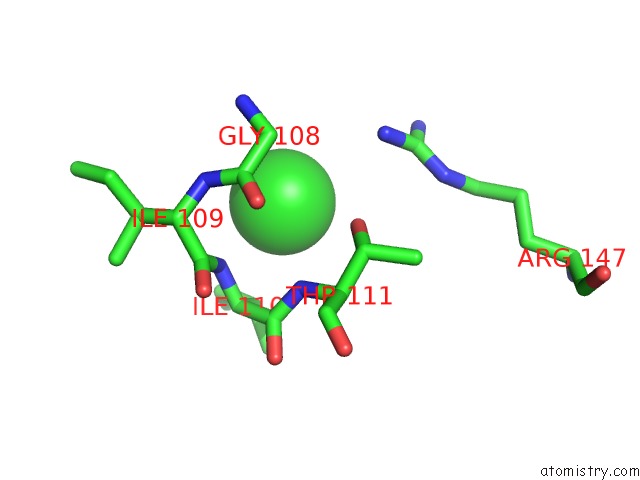
Chlorine binding site 4 out
of 6 in the Crystal Structure of Probable Acetyltransferase
 Mono view
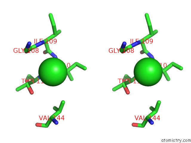
 Stereo pair view
|
A full contact list of Chlorine with other atoms in the Cl binding
site number 4 of Crystal Structure of Probable Acetyltransferase within 5.0Å range:
probe
|
atom
|
residue
|
distance (Å)
|
B
|
Occ
|
D:Cl184
b:81.5
occ:0.75
|
CB
|
D:ILE110
|
4.1
|
26.6
|
1.0
|
N
|
D:ILE110
|
4.1
|
27.9
|
1.0
|
N
|
D:THR111
|
4.2
|
29.2
|
1.0
|
CA
|
D:GLY108
|
4.2
|
30.1
|
1.0
|
OG1
|
D:THR111
|
4.4
|
32.5
|
1.0
|
N
|
D:ILE109
|
4.4
|
28.9
|
1.0
|
CG2
|
D:VAL144
|
4.4
|
24.7
|
1.0
|
N
|
D:GLY108
|
4.5
|
30.1
|
1.0
|
C
|
D:GLY108
|
4.5
|
29.4
|
1.0
|
CA
|
D:ILE110
|
4.5
|
27.4
|
1.0
|
CG1
|
D:ILE110
|
4.7
|
22.9
|
1.0
|
CB
|
D:THR111
|
4.8
|
30.6
|
1.0
|
CD1
|
D:ILE110
|
4.8
|
14.7
|
1.0
|
C
|
D:ILE110
|
4.9
|
28.3
|
1.0
|
|
Chlorine binding site 5 out
of 6 in 1nsl
Go back to
Chlorine Binding Sites List in 1nsl
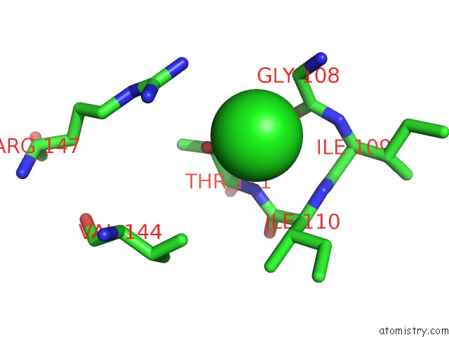
Chlorine binding site 5 out
of 6 in the Crystal Structure of Probable Acetyltransferase
 Mono view
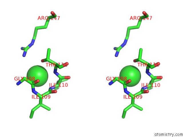
 Stereo pair view
|
A full contact list of Chlorine with other atoms in the Cl binding
site number 5 of Crystal Structure of Probable Acetyltransferase within 5.0Å range:
probe
|
atom
|
residue
|
distance (Å)
|
B
|
Occ
|
E:Cl184
b:84.6
occ:0.60
|
N
|
E:ILE110
|
3.2
|
28.6
|
1.0
|
OG1
|
E:THR111
|
3.3
|
29.7
|
1.0
|
CB
|
E:ILE110
|
3.5
|
28.8
|
1.0
|
CA
|
E:GLY108
|
3.6
|
30.8
|
1.0
|
C
|
E:GLY108
|
3.6
|
30.2
|
1.0
|
N
|
E:ILE109
|
3.7
|
29.3
|
1.0
|
N
|
E:THR111
|
3.7
|
29.3
|
1.0
|
CG1
|
E:ILE110
|
3.8
|
27.7
|
1.0
|
CA
|
E:ILE110
|
3.8
|
29.4
|
1.0
|
N
|
E:GLY108
|
4.1
|
31.4
|
1.0
|
C
|
E:ILE109
|
4.1
|
28.8
|
1.0
|
O
|
E:GLY108
|
4.2
|
30.7
|
1.0
|
C
|
E:ILE110
|
4.3
|
29.9
|
1.0
|
NH2
|
E:ARG147
|
4.3
|
34.1
|
1.0
|
CA
|
E:ILE109
|
4.4
|
28.8
|
1.0
|
CB
|
E:THR111
|
4.5
|
30.0
|
1.0
|
CA
|
E:THR111
|
4.7
|
29.6
|
1.0
|
CD1
|
E:ILE110
|
4.7
|
25.9
|
1.0
|
CZ
|
E:ARG147
|
4.8
|
36.7
|
1.0
|
CG2
|
E:ILE110
|
4.9
|
28.6
|
1.0
|
CB
|
E:ILE109
|
4.9
|
28.5
|
1.0
|
|
Chlorine binding site 6 out
of 6 in 1nsl
Go back to
Chlorine Binding Sites List in 1nsl
Chlorine binding site 6 out
of 6 in the Crystal Structure of Probable Acetyltransferase
 Mono view
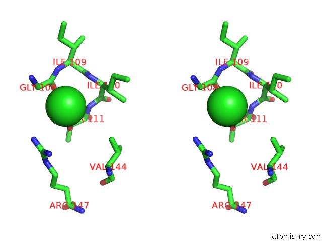
 Stereo pair view
|
A full contact list of Chlorine with other atoms in the Cl binding
site number 6 of Crystal Structure of Probable Acetyltransferase within 5.0Å range:
probe
|
atom
|
residue
|
distance (Å)
|
B
|
Occ
|
F:Cl184
b:86.7
occ:1.00
|
OG1
|
F:THR111
|
2.9
|
30.3
|
1.0
|
CA
|
F:GLY108
|
3.4
|
33.1
|
1.0
|
C
|
F:GLY108
|
3.7
|
31.9
|
1.0
|
N
|
F:THR111
|
3.7
|
31.1
|
1.0
|
N
|
F:GLY108
|
3.9
|
33.4
|
1.0
|
CB
|
F:THR111
|
4.0
|
31.5
|
1.0
|
NH2
|
F:ARG147
|
4.0
|
43.6
|
1.0
|
N
|
F:ILE110
|
4.0
|
30.5
|
1.0
|
N
|
F:ILE109
|
4.0
|
31.3
|
1.0
|
O
|
F:GLY108
|
4.2
|
32.0
|
1.0
|
CB
|
F:ILE110
|
4.2
|
30.6
|
1.0
|
CZ
|
F:ARG147
|
4.4
|
42.9
|
1.0
|
NH1
|
F:ARG147
|
4.4
|
43.0
|
1.0
|
CA
|
F:THR111
|
4.5
|
31.4
|
1.0
|
CA
|
F:ILE110
|
4.5
|
31.3
|
1.0
|
C
|
F:ILE110
|
4.6
|
31.5
|
1.0
|
C
|
F:ILE109
|
4.8
|
30.9
|
1.0
|
CG2
|
F:VAL144
|
4.9
|
24.6
|
1.0
|
CG1
|
F:ILE110
|
4.9
|
29.6
|
1.0
|
CA
|
F:ILE109
|
5.0
|
30.7
|
1.0
|
|
Reference:
J.S.Brunzelle,
R.Wu,
S.V.Korolev,
F.R.Collart,
A.Joachimiak,
W.F.Anderson.
Crystal Structure of Bacillus Subtilis Ydaf Protein: A Putative Ribosomal N-Acetyltransferase Proteins V. 57 850 2004.
ISSN: ISSN 0887-3585
PubMed: 15468321
DOI: 10.1002/PROT.10601
Page generated: Sat Jul 20 00:37:12 2024
|